
|
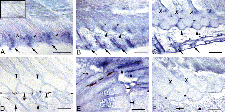
Fig. 4 Expression of bmp2b in the median fins of late-stage zebrafish. Arrows mark fin rays, arrowheads mark radials/hypurals. Black patches are melanophores. Anterior is to the left and dorsal to the top. Presumptive fin ray tissue within the anal fin fold expresses bmp2b, distal to the muscle buds () (5.2 mm NL) (A). Radials, not in this plane of section, but see inset, do not express bmp2b at this stage (A). Fin ray expression is maintained and a new domain of expression in the ZS (*) of the anal and dorsal fins is observed (5.5 mm) (B). A third domain of bmp2b expression in the maturing and hypertrophic chondrocytes (X) of the anal and dorsal fin radials develops in larger specimens; fin ray and ZS expression is maintained (6.0 mm) (C). By 7.3 mm, expression in the fin rays, ZS, and hypertrophic chondrocytes of the anal and dorsal fins is no longer detected (D). bmp2b expression is observed in caudal fin rays and in hypertrophic chondrocytes of the caudal fin supports, but not in mesenchyme or proliferative chondrocytes (6.6 mm) (E). No cells are distinctly labeled by bmp2b sense probe (6.6 mm) (F). Scale bars equal 0.05 mm.
Reprinted from Gene expression patterns : GEP, 5(2), Crotwell, P.L., Sommervold, A.R., and Mabee, P.M., Expression of bmp2a and bmp2b in late-stage zebrafish median fin development, 291-296, Copyright (2004) with permission from Elsevier. Full text @ Gene Expr. Patterns