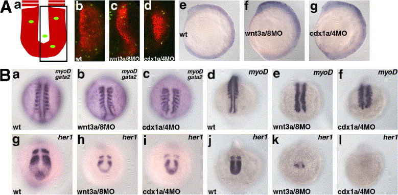
Fig. 8 The Wnt-Cdx cascade is involved in morphogenesis of the posterior body. (A) Cell proliferation and cell death was not strongly impaired in the in the wnt3a/wnt8 (c, f) and cdx1a/cdx4 morphant embryos (d, g) (b, e, control embryos). The morphant embryos displayed the Class III phenotype. The embryos were fixed at 17 hpf and stained with the paraxial mesoderm marker papc probe (red) and with anti-phosphohistone H3 antibodies (green). (Aa) Schematic representation of the stained embryos and (Ab–d) confocal images of representative embryos. Note that the number of phosphoH3-positive cells was not significantly different among the wild-type, wnt3a/wnt8-morphant, and cdx1a/cdx4 morphant embryos. (Ae–g) The embryos were fixed at 13 hpf and subjected to Tunel staining. (B) Impairment of the posterior somite formation. Expression of myoD at 13 hpf (a–c, gata2 was also stained) and 17 hpf (d–f). Expression of her1 at 13 hpf (g–i) and 17 hpf (j–l). (a–f) dorsal views and (g–l) posterior views. her1 expression was reduced but displayed normal oscillation in the wnt3a/wnt8 and cdx1a/cdx4 morphant embryos at the early segmentation stage, which had normal anterior somites. At the mid-to-late segmentation periods, her1 expression was strongly reduced or abrogated in the wnt3a/wnt8 and cdx1a/cdx4 morphant embryos, and segmentation stopped for the posterior somites.
Reprinted from Developmental Biology, 279(1), Shimizu, T., Bae, Y.K., Muraoka, O., and Hibi, M., Interaction of Wnt and caudal-related genes in zebrafish posterior body formation, 125-141, Copyright (2005) with permission from Elsevier. Full text @ Dev. Biol.

