
IMAGE
Fig. 4
- ID
- ZDB-IMAGE-071001-101
- Genes
- Publication
- Te Velthuis et al., 2007 - Gene expression patterns of the ALP family during zebrafish development
- All Figures
- Figures for Te Velthuis et al., 2007
Image
|
Figure Caption
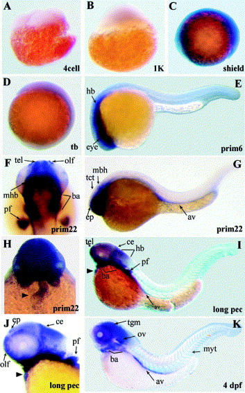
Fig. 4 Expression pattern of the zebrafish elfin gene by in situ hybridization. Picture of the in situ experiments with zebrafish elfin specific probes show staining patterns in the regions of the central nervous system (telencephalon (tel), olfactory placode (olf), cerebellum (ce) and hindbrain (hb)), the pectoral fins (pf), branchial arches (ba) and the heart (black arrowhead). Further expression was observed in the axial vasculature (av), epiphysis (epi), midbrain-hindbrain boundary (mhb), myotomes (myt) and the otic vesicle (ov).
Figure Data
Acknowledgments
This image is the copyrighted work of the attributed author or publisher, and
ZFIN has permission only to display this image to its users.
Additional permissions should be obtained from the applicable author or publisher of the image.
Reprinted from Gene expression patterns : GEP, 7(3), Te Velthuis, A.J., Ott, E.B., Marques, I.J., and Bagowski, C.P., Gene expression patterns of the ALP family during zebrafish development, 297-305, Copyright (2007) with permission from Elsevier. Full text @ Gene Expr. Patterns