
IMAGE
Fig. 6
- ID
- ZDB-IMAGE-070925-98
- Genes
- Publication
- Albertson et al., 2007 - Fgf8 haploinsufficiency results in distinct craniofacial defects in adult zebrafish
- All Figures
- Figures for Albertson et al., 2007
Image
|
Figure Caption
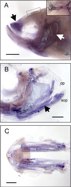
Fig. 6 Fig. 6. Fgf8 mRNA is expressed in distinct anatomical regions of the mature craniofacial skeleton. (A) A 6-month-old wild-type zebrafish head showing fgf8 expression in the upper jaw apparatus (black arrow), operculum (white arrow), and sagittal suture (inset). Scale bar equals 1 mm. (B) The dissected operculum shows fgf8 expression throughout the subopercle (sop) and along the proximal–distal surface of the opercle (op) (black arrow). Scale bar = 500 μm. (C) Expression of fgf8 is also observed along the ventral surface of the mandible. Scale bar = 500 μm.
Figure Data
Acknowledgments
This image is the copyrighted work of the attributed author or publisher, and
ZFIN has permission only to display this image to its users.
Additional permissions should be obtained from the applicable author or publisher of the image.
Reprinted from Developmental Biology, 306(2), Albertson, R.C., and Yelick, P.C., Fgf8 haploinsufficiency results in distinct craniofacial defects in adult zebrafish, 505-515, Copyright (2007) with permission from Elsevier. Full text @ Dev. Biol.