
|
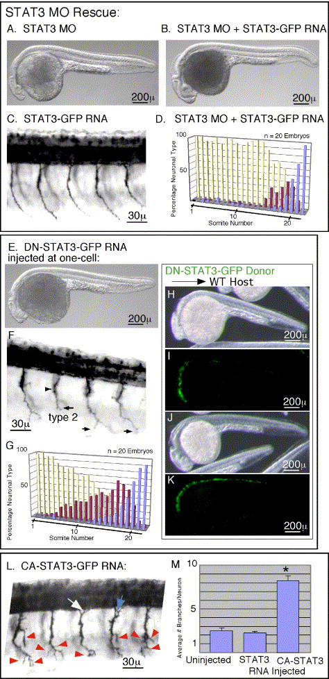
Fig. 3 Morpholino defects can be rescued, while Dominant negative and Constitutively Active versions differentially alter Pathfinding. In panel F, black arrowheads mark the choice point and black arrows mark the most ventral extension of the CaP motoneuron axon. In panel L, red arrowheads mark axon branches. White and blue arrows in panel L are mentioned in the Results section on mosaic analysis. (A–D) Rescue of Morpholino induced pathfinding defects. (A) Whole body image showing foreshortened anterior axis with subsequent fore and mid-brain defects. (B) Whole body image showing rescue of the anterior axis by co-injection of RNA encoding wild-type STAT3 with the STAT3 MO. (C) Primary motoneuron projections in an embryo injected with RNA encoding wild-type STAT3, showing no affect upon pathfinding. (D) Quantitation of CaP neural types showing rescue of pathfinding defects by co-injection of MO with RNA encoding wild-type STAT3. (E–G) Analysis of embryos expressing DN-STAT3. (E) Whole body image showing foreshortened anterior axis with subsequent fore and mid-brain defects. (F) Primary motoneuron projections showing forestalled ventral CaP axon projections. (G) Quantitation of CaP neural types showing stalled type 2 and 3 neurons in rostral as well as caudal somites. (H–K) Confirmation that DN-STAT3 inhibits endogenous signaling by demonstration of nonautonomous cell migration defects. (H) Whole body image of a wild-type embryo showing foreshortened anterior axis with subsequent fore and mid-brain defects due to transplantation of DN-STAT3 expressing cells populating anterior axial mesendoderm. Because these cells contribute to precordal mesendoderm, they inhibited migration of neighboring cells along the anterior axis. This noncell autonomous affect during gastrulation is a characteristic of STAT3 signaling. (I) GFP fluorescence of donor cells populating anterior axial mesendoderm of embryo shown in panel H. (J) Whole body image of a wild-type embryo showing normal anterior axis because DN-STAT3 expressing cells did not populate the precordal plate. (K) The donor cells, shown by GFP fluorescence, positioned more posterior to those in panels H and I, and thus not inhibiting migration of neighboring blastomeres. (L and M) Constitutive activation of STAT3 leads to exuberant branching. (L) Primary motoneuron projections showing extra branches on the CaP motoneuron axon. (M) Quantitation of branch numbers. Asterisk marks statistical significance (t test, P < 0.05). In all images, rostral is to the left, and dorsal is up. Embryos are at 27 h post-fertilization.
Reprinted from Developmental Biology, 296(1), Conway, G., STAT3-dependent pathfinding and control of axonal branching and target selection, 119-136, Copyright (2006) with permission from Elsevier. Full text @ Dev. Biol.