
|
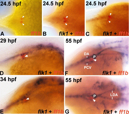
Fig. 1 Spatial relationship between interrenal tissue and endothelium during early zebrafish development. Developing wild type embryos at 24.5 hpf (A–C), 29 hpf (D), 34 hpf (E) and 55 hpf (F, G) were subject to either single ISH for ff1b (red) (A), or double ISH for flk1 (dark blue) and ff1b (red) (B–G). Panels A, B and G are dorsal views, while panels C–F are dorsolateral views of the trunk region. All panels are oriented with anterior to the right. The tight association between interrenal tissue and endothelium initiates before the convergence of interrenal primordia. DA, dorsal aorta; PCV, posterior cardinal vein; P, pronephros; LDA, lateral dorsal aortae. White arrowheads indicate the interrenal tissues.
Reprinted from Developmental Biology, 297(1), Liu, Y.W., and Guo, L., Endothelium is required for the promotion of interrenal morphogenetic movement during early zebrafish development, 44-58, Copyright (2006) with permission from Elsevier. Full text @ Dev. Biol.