
|
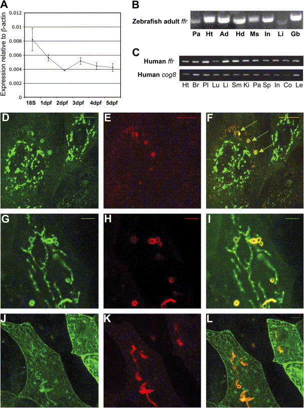
Fig. 6 ffr expression and protein localization A) Developmental profile of ffr expression. mRNA levels were determined by a zebrafish oligo microarray. Normalized data are expressed relative to β-actin and from 18 somite to 5 dpf stages. Data represent mean ± SEM, (n = 3–6 at each stage). B) ffr is widely expressed in the adult zebrafish. Expression of ffr was determined by RT-PCR. Tissues: Pa, pancreas; Ht, heart; Ad, adipose tissue; Hd: head; Ms; muscle; In: intestine; Li: liver; Gb, gall bladder. C) Human ffr and COG8 are widely expressed. Expression was assayed by RT-PCR. Tissues: Ht, heart; Br, brain; Pl, placenta; Lu, lung; Li, liver; SM, skeletal muscle; Ki, kidney; Pa, pancreas; Sp, spleen; In, small intestine; Co, colon; Le, leucocyte. D–F) Ffr and COG8 partially colocalize in the Golgi of zebrafish blastomeres. Embryos were coinjected with ffr-gfp mRNA and cog8-mrfp mRNA at the 1- to 4-cell stage. D) Expression of Ffr-GFP in the perinuclear region. E) COG8-mRFP localizes primarily to the peripheral Golgi. F) Merged image of (D) and (E). Arrowheads indicate colocalization of COG8 and Ffr. G–I) Wild-type (in green) and mutant (in red) Ffr partially colocalize in the cells. (G) Expression of ffrWt-GFP. (H) Expression of ffr mut-mRFP. (I) Merged image of (G) and (H). Yellow indicates colocalization. J–L) Mutant Ffr (in red) and clathrin light chain (in green) colocalization. (J) TGN marker (Clathrin, GFP; pClGFP) (Gaidarov et al., 1999) expression in a single enveloping layer cell. (K) ffr mut-mRFP expression in a single cell. (L) Merged image of (J) and (K). Yellow indicates truncated Ffr mutant localized in the TGN. The scale bar represents 5 μm.
Reprinted from Cell Metabolism, 3(4), Ho, S.Y., Lorent, K., Pack, M., and Farber, S.A., Zebrafish fat-free is required for intestinal lipid absorption and Golgi apparatus structure, 289-300, Copyright (2006) with permission from Elsevier. Full text @ Cell Metab.