
IMAGE
Fig. 5
- ID
- ZDB-IMAGE-070917-92
- Genes
- Publication
- Tendeng et al., 2006 - Cloning and embryonic expression of five distinct sfrp genes in the zebrafish Danio rerio
- All Figures
- Figures for Tendeng et al., 2006
Image
|
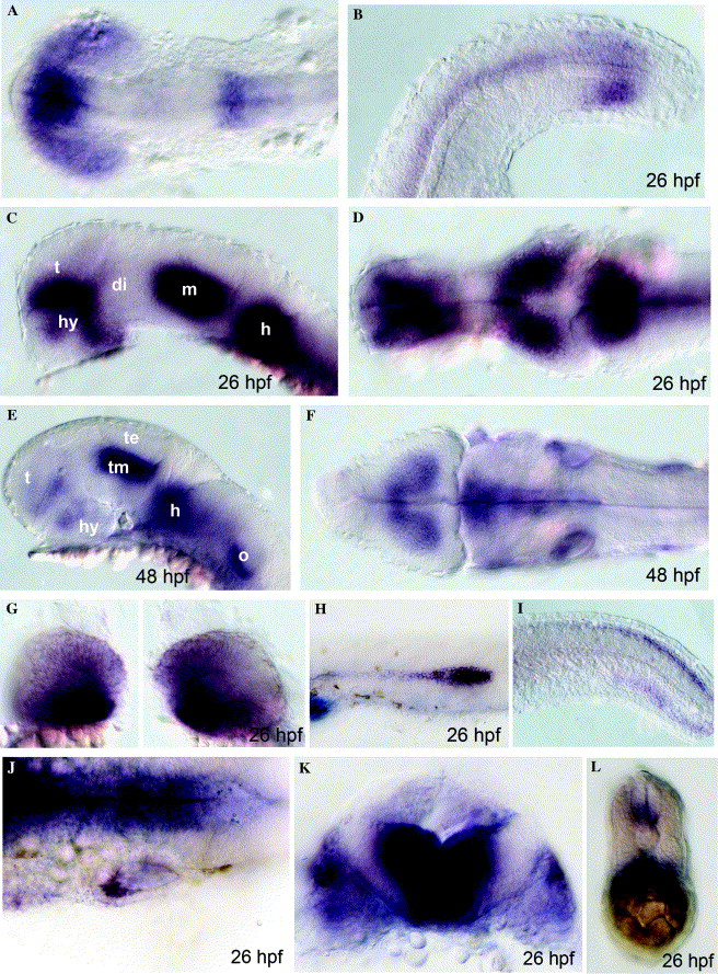
Figure Caption
Fig. 5 Later expression profiles of zebrafish sfrp1a. Dorsal (A, D, F and J), lateral (B, C, E and G-I) or transversal (K and L) views of zebrafish embryos hybridized with sfrp1a antisense probe. Abbreviations: di, diencephalon; h, hindbrain; hy, hypothalamus; m, midbrain; o, otic vesicle; t, telencephalon; te, optic tectum; tm, tegmentum.
Figure Data
Acknowledgments
This image is the copyrighted work of the attributed author or publisher, and
ZFIN has permission only to display this image to its users.
Additional permissions should be obtained from the applicable author or publisher of the image.
Reprinted from Gene expression patterns : GEP, 6(8), Tendeng, C., and Houart, C., Cloning and embryonic expression of five distinct sfrp genes in the zebrafish Danio rerio, 761-771, Copyright (2006) with permission from Elsevier. Full text @ Gene Expr. Patterns