
|
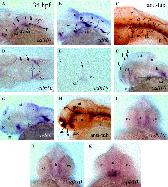
Fig. 5 cdh10 expression in 34 hpf embryos (A, D-F, and I-K) compared to cdh6 expression (B and G) and anti-acetylated tubulin staining (C and H). Panels (A-C and F-H) are lateral views of whole mount embryo heads with anterior to the left and dorsal up. (D) A dorsal view (anterior to the left) of a deyolked embryo. (E) A parasagittal section of the hindbrain (anterior to the left and dorsal up). (I)-(K) Frontal views (dorsal up) focusing on planes indicated by three correspondingly labeled arrows in (F). The arrowhead, long and short arrows in (A), (D), and (E) point to the same cdh10 expressing cell clusters. Abbreviations: ad, anterodorsal lateral line ganglion; av, anteroventral lateral line ganglion; hy, hypothalamus; m/v, medial lateral line/vagus ganglion, r1-r6, rhombomeres 1-6; and t, trigeminal ganglion. The remaining abbreviations are the same as in Fig. 4.
Reprinted from Gene expression patterns : GEP, 74(6), Liu, Q., Duff, R.J., Liu, B., Wilson, A.L., Babb-Clendenon, S.G., Francl, J., and Marrs, J.A., Expression of cadherin10, a type II classic cadherin gene, in the nervous system of the embryonic zebrafish, 1016-1025, Copyright (2006) with permission from Elsevier. Full text @ Gene Expr. Patterns