
|
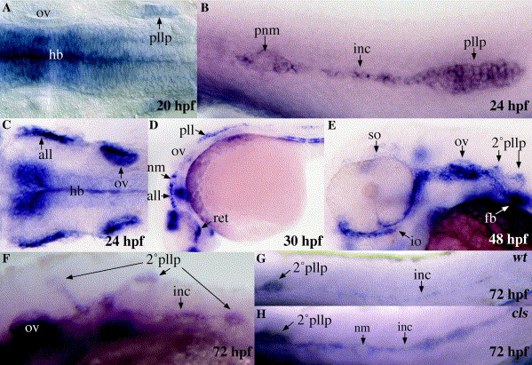
Fig. 4 Analysis of sfrp1a expression in the developing lateral line. Whole mount in situ hybridisation was performed with an antisense RNA probe for sfrp1a at the indicated stages (A–H). A wild-type embryo is compared to a cls mutant embryo (G and H). Embryos are presented in whole mount with anterior to the left, in dorsal (A and C) or lateral view (B and D–H). all, anterior lateral line; fb, fin-bud; hb, hindbrain; inc, interneuromast cells; io, infraorbital lateral line; nm, neuromast; ov, otic vesicle; pll, posterior lateral line; pllp, posterior lateral line placode; pnm, proneuromast; ret, retina; so, supraorbital lateral line; 2°pllp, secondary posterior lateral line primordium.
Reprinted from Gene expression patterns : GEP, 6(8), Pezeron, G., Anselme, I., Laplante, M., Ellingsen, S., Becker, T.S., Rosa, F.M., Charnay, P., Schneider-Maunoury, S., Mourrain, P., and Ghislain, J., Duplicate sfrp1 genes in zebrafish: sfrp1a is dynamically expressed in the developing central nervous system, gut and lateral line, 835-842, Copyright (2006) with permission from Elsevier. Full text @ Gene Expr. Patterns