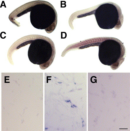
Fig. 2 Detection of hsp27 gene expression by in situ hybridization. In situ hybridization was conducted using embryos fixed 24 hpf. Embryos shown are: (A) processed without probe, (B) pretreated with DNAse I and RNAse prior to incubation with antisense, Hsp27 mRNA specific probe, (C) incubated with a sense control probe, and (D) incubated with antisense, Hsp27 mRNA specific probe. Hsp27 mRNA is detected unevenly in trunk and tail myotomes. NIH-3T3 murine fibroblasts were transiently transfected with an expression vector for an EGFP-zebrafish Hsp27 fusion protein. Cell cultures were processed using a sense control probe (E), antisense probe (F) or pretreated with DNAse and RNAse before incubation with antisense probe (G). Bar =200 μm (A–D), 100 μm (E–G).
Reprinted from Gene expression patterns : GEP, 6(2), Mao, L., and Shelden, E.A., Developmentally regulated gene expression of the small heat shock protein Hsp27 in zebrafish embryos, 127-133, Copyright (2006) with permission from Elsevier. Full text @ Gene Expr. Patterns

