
|
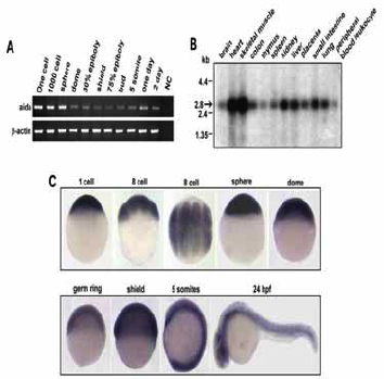
Fig. S5 Temporal and spatial expression of aida. (A) Developmental expression pattern of aida in zebrafish was determined by reverese transcription polymerase chain reaction (RT-PCR). RNA sample from one-cell embryos with no addition of reverse transcriptase was used as negative control Zebrafish β-actin was the loading control. (B) Northern blot with poly(A)+ RNA isolated from various tissues of human, indicated on the top, was hybridized with 32P-labelled human AIDA DNA probe. the band representing Aida was indicated by an arrow. (C) Spatiotemporal expression of zebrafish aida was detected by whole-mount in situ hybridization at different stages at indicated stages. Note that aida is ubiquitously expressed throughout these stages.
Reprinted from Developmental Cell, 13(2), Rui, Y., Xu, Z., Xiong, B., Cao, Y., Lin, S., Zhang, M., Chan, S.C., Luo, W., Han, Y., Lu, Z., Ye, Z., Zhou, H.M., Han, J., Meng, A., and Lin, S.C., A beta-Catenin-Independent Dorsalization Pathway Activated by Axin/JNK Signaling and Antagonized by Aida, 268-282, Copyright (2007) with permission from Elsevier. Full text @ Dev. Cell