
IMAGE
Fig. 3
- ID
- ZDB-IMAGE-070822-12
- Genes
- Publication
- Shi et al., 2006 - Zebrafish foxe3: Roles in ocular lens morphogenesis through interaction with pitx3
- All Figures
- Figures for Shi et al., 2006
Image
|
Figure Caption
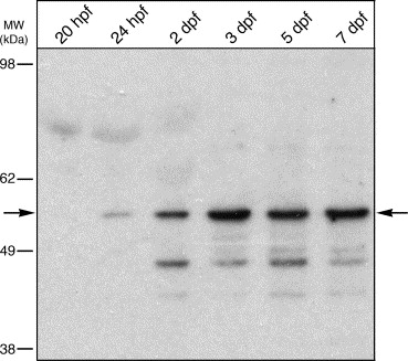
Fig. 3 Temporal expression of Foxe3 protein. Immunoblot detection of Foxe3 in extracts from 20 and 24 hpf whole embryos and heads at 2, 3, 5 and 7 dpf. The Foxe3 protein is barely detected at 20 and 24 hpf (arrow). The expression level increases through 7 dpf compared to the earlier time points. Relative location of molecular weight markers are shown to the left (in kDa).
Figure Data
Acknowledgments
This image is the copyrighted work of the attributed author or publisher, and
ZFIN has permission only to display this image to its users.
Additional permissions should be obtained from the applicable author or publisher of the image.
Reprinted from Mechanisms of Development, 123(10), Shi, X., Luo, Y., Howley, S., Dzialo, A., Foley, S., Hyde, D.R., and Vihtelic, T.S., Zebrafish foxe3: Roles in ocular lens morphogenesis through interaction with pitx3, 761-782, Copyright (2006) with permission from Elsevier. Full text @ Mech. Dev.