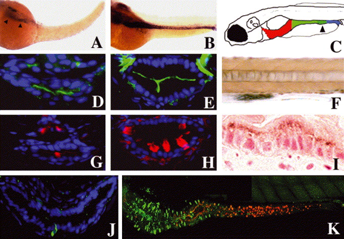
Fig. 5 Smooth muscle progenitors and epithelial differentiation: (A,B) Whole-mount RNA in situ showing intestinal smooth muscle myosin heavy chain expression at 50 (A, arrowheads) and 58 hpf (B). (C) Cartoon depicting anterior (red), mid (green) and posterior (blue) segments of the 5 dpf larval intestine. Distal region of the mid intestine that contains specialized enterocytes is stippled (arrowhead). (D,E,G,H) Cross-sections through the larval mid intestine. (D,E) Enterocytes contain the NaPi transporter within their apical cell membrane. NaPi+ enterocytes are first identified at 74 hpf (D); NaPi levels are pronounced at 120 hpf (E). (G) Goblet cell mucin, revealed here by rhodamine-dextran labeled WGA, is first evident at 78 hpf. (H) Mucin is much more abundant at 120 hpf and is easily recognized in histological specimens at this stage (not shown). (J) Cross-section through the anterior intestine of a 96 hpf larva. Pancreatic polypeptide+ enteroendocrine cells are first identified at this stage. Note slender apical cytoplasm characteristic of these cells. (K) Lateral view of the 120 hpf intestine showing regionalized distribution of differentiated epithelial cells. Pancreatic polypeptide containing enteroendocrine cells (green) are restricted to the anterior intestine (intestinal bulb) whereas goblet cells identified with WGA (red) are restricted to the mid intestine. NaPi+ enterocytes are present throughout the anterior and mid intestine, but not the posterior intestine (not shown). (F) Lateral view of a portion of the mid intestine from a 96 hpf larva that has ingested horseradish peroxidase protein (HRP). Following pinocytosis, HRP can be detected histochemically within the apical cytoplasm of specialized enterocytes of the mid intestine (segment 2) that also express NaPi (not shown). (I) Sagittal histological section through the mid intestine of the larva shown in (F), reveals HRP within the apical enterocyte cytoplasm.
Reprinted from Mechanisms of Development, 122(2), Wallace, K.N., Akhter, S., Smith, E.M., Lorent, K., and Pack, M., Intestinal growth and differentiation in zebrafish, 157-73, Copyright (2005) with permission from Elsevier. Full text @ Mech. Dev.

