
Fig. 5
- ID
- ZDB-IMAGE-070815-19
- Genes
- Publication
- Zhao et al., 2007 - Genetic defects of pronephric cilia in zebrafish
- All Figures
- Figures for Zhao et al., 2007
|
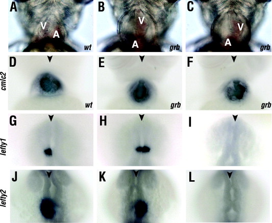
Fig. 5 Left–right asymmetry phenotype in grbtm304 mutant embryos. (A–C) Ventral views of wild-type and grbtm304 embryos at 72 hpf. Wild-type animals display normal heart looping phenotype (A). Heart looping is frequently absent (B) or inverted (C) in grbtm304 mutant embryos. (D–F) In situ hybridization staining for a heart marker, cmlc2, at 3 dpf. Normal heart orientation in the wild type is shown in (D). The absence of obvious heart looping (E) or reverse looping (F) in mutant embryos. (G–I) The expression of lefty1 in embryos collected from crosses between grbtm304/+ heterozygotes. The lefty1 transcript is expressed on the left side of neural tube in the majority of embryos (G). The lefty1 expression domain extends to both sides (H) or is absent (I) in some embryos. (J, K, and L) The expression of lefty2 in embryos collected from crosses between grbtm304/+ heterozygotes. (J) The normal expression of lefty2 transcript on the left side of the lateral plate mesoderm. The lefty2 expression domain shifts to the right side (K) or is absent (L) in some embryos. A, atrium; V, ventricle. In all panels anterior is up. Arrowhead indicates the midline.
Reprinted from Mechanisms of Development, 124(7-8), Zhao, C., and Malicki, J., Genetic defects of pronephric cilia in zebrafish, 605-616, Copyright (2007) with permission from Elsevier. Full text @ Mech. Dev.