
Fig. 7
- ID
- ZDB-IMAGE-070801-7
- Publication
- Jin et al., 2007 - A transgene-assisted genetic screen identifies essential regulators of vascular development in vertebrate embryos
- All Figures
- Figures for Jin et al., 2007
|
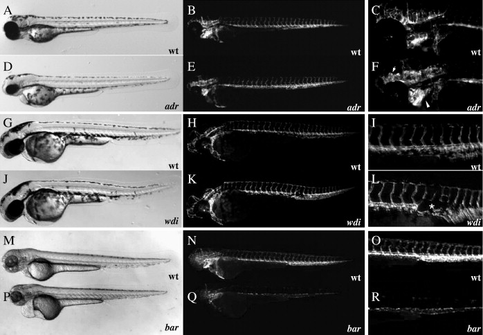
Fig. 7 Mutations affecting vascular maintenance and integrity. (A–R) Bright-field (A, D, G, J, M, and P) and epifluorescent (B, C, E, F, H, I, K, L, N, O, Q, and R) micrographs of Tg(flk1:EGFP)s843adrs277 mutant larvae (D–F) and wild-type siblings (A–C), wdis631 mutant larvae (J–L) and wild-type siblings (G–I), and bars847 mutant larvae (P–R) and wild-type siblings (M–O) at 72 hpf. Affected areas within epifluorescent micrographs are magnified and shown in panels C, F, I, L, O, and R. Note the massive vascular dilation throughout the head vasculature in adrs277 mutant larvae (F arrowheads), regression of the dorsal aorta in wdis631 mutant larvae (asterisk in panels L; K, L), and apparently disappearing endothelial cells in bars847 mutant larvae (Q, R).
Reprinted from Developmental Biology, 307(1), Jin, S.W., Herzog, W., Santoro, M.M., Mitchell, T.S., Frantsve, J., Jungblut, B., Beis, D., Scott, I.C., D'Amico, L.A., Ober, E.A., Verkade, H., Field, H.A., Chi, N.C., Wehman, A.M., Baier, H., and Stainier, D.Y., A transgene-assisted genetic screen identifies essential regulators of vascular development in vertebrate embryos, 29-42, Copyright (2007) with permission from Elsevier. Full text @ Dev. Biol.