
|
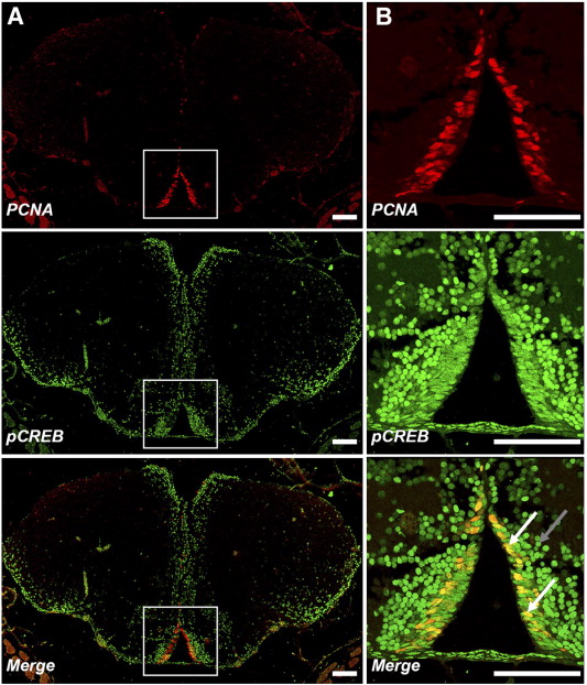
Fig. 4 Proliferating neural cells express pCREB. Double-labelling immunofluorescence shows actively-proliferating cells in PZ2 are pCREB+. Coronal sections of adult zebrafish anterior telencephalon double-stained for PCNA (red) and pCREB (green) show most PCNA + cells lining the walls of the ventral diencephalic ventricles are pCREB+ (white box; x100 A and x400; B). Additional pCREB+ cells extending laterally into the telencephalon correspond to regions of migrating neuroblasts. Note differences in morphology between flattened, elongated pCREB+/PCNA + cells adjacent to the ventricular wall (white arrows) compared to smaller, round pCREB+/PCNA- cells (grey arrow) which have migrated tangentially into the telencephalon proper from the ventricular proliferation zone.
Reprinted from Developmental Biology, 307(1), Dworkin, S., Heath, J.K., Dejong-Curtain, T.A., Hogan, B.M., Lieschke, G.J., Malaterre, J., Ramsay, R.G., and Mantamadiotis, T., CREB activity modulates neural cell proliferation, midbrain-hindbrain organization and patterning in zebrafish, 127-141, Copyright (2007) with permission from Elsevier. Full text @ Dev. Biol.