
Fig. 4
- ID
- ZDB-IMAGE-070620-38
- Publication
- Holtzman et al., 2007 - Endocardium is necessary for cardiomyocyte movement during heart tube assembly
- All Figures
- Figures for Holtzman et al., 2007
|
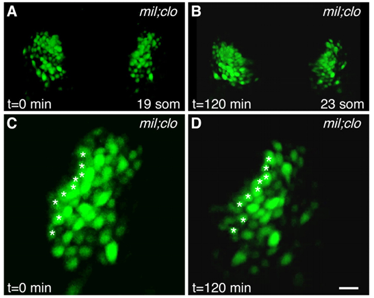
Fig. 4 Endocardium directs cardiomyocyte movement, even in the absence of initial medial movement. (A-D) Selected images from a time-lapse of cardiac fusion in a mil;clo double-mutant zebrafish embryo expressing Tg(cmlc2:egfp) (see Movie 4 in the supplementary material), exhibiting cardiac morphology at the (A) 19-somite and (B) 23-somite stages. Dorsal views, anterior to the top. (C,D) Paths traveled by cardiomyocytes in the right lateral heart field during the entire time-lapse (asterisks as described in Fig. 1). Images in C,D are three times the magnification of those in A,B. Scale bar: 20 μm. Example shown is representative of four heart fields analyzed. Cardiomyocytes in mil;clo double mutants exhibit no net directed movement.