
Fig. S4
- ID
- ZDB-IMAGE-070606-3
- Genes
- Publication
- Nyholm et al., 2007 - The zebrafish zic2a-zic5 gene pair acts downstream of canonical Wnt signaling to control cell proliferation in the developing tectum
- All Figures
- Figures for Nyholm et al., 2007
|
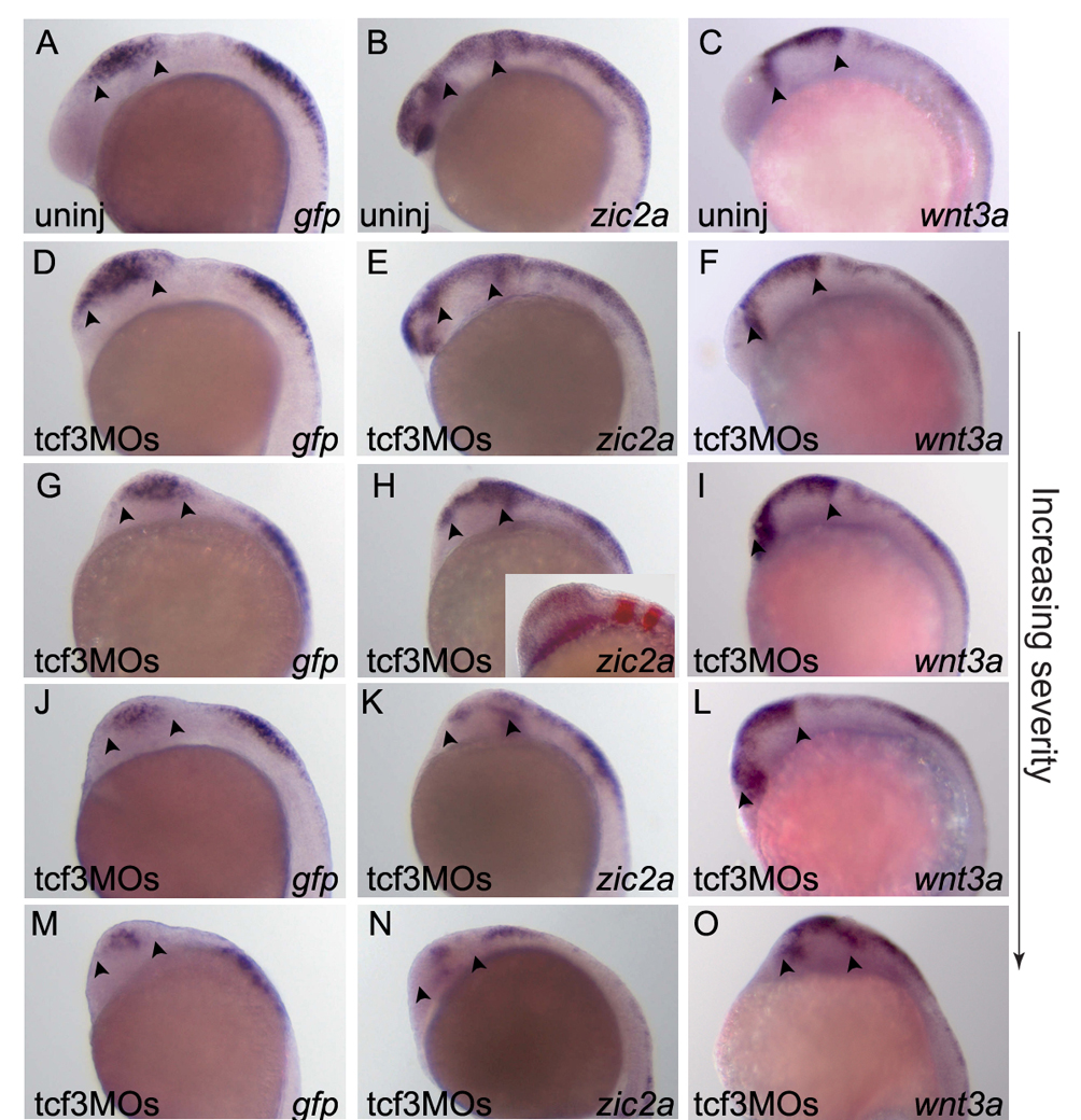
Fig. S4 Tcf3 reduction affects midbrain size. (A-C) Uninjected control embryos showing normal zic2aD5:gfp (A), zic2a (B) and wnt3a (C) expression at 18-somites. (D-O) Embryos injected with tcf3 MOs and assayed for zic2aD5:gfp (G,J,M), zic2a (H,K,N), or wnt3a (I,L,O) expression at 18-somites. Knockdown of Tcf3 resulted in expanded expression of all markers in the midbrain at low MO concentrations. This is most likely due to the previously documented overall midbrain expansion (D-I). At higher MO concentrations, zic2aD5:gfp and zic2a were reduced in midbrain (J,M,K,N), whereas wnt3a expression remained strong (L,O). Embryos are shown in lateral view, anterior to the left. Arrowheads mark anterior and posterior limits of the midbrain.