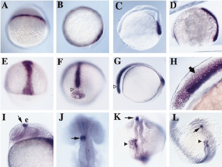
Fig. 4 Whole mount in situ hybridization of ndr2 expression. (A) Dome stage, lateral view. (B) 30% epiboly, animal view; ndr2 is expressed at the blastoderm margin. (C) Shield stage, lateral view (dorsal to the right); ndr2 expression is restricted to the hypoblast layer of the shield. (D) 80% epiboly, lateral view, and (E) 70% epiboly, dorsal view; ndr2 expression is seen in involuting hypoblast. (F–H) Bud stage; anterior view (F), lateral view with anterior to the left (G), higher magnification lateral view (H). ndr2 is present in an anterior domain including the prechordal plate, anterior notochord, and ventral neuroectoderm (arrow); open triangles mark a gap in the anterior domain, asterisks indicate the polster, and a dotted line traces Brachet’s cleft separating anterior mesoderm and neurectoderm. ndr2 also is expressed in the tail bud (G). (I–L) 20-somite stage. Lateral view (I; anterior to the left), and dorsal view of the head (J; anterior to the top); ndr2 is expressed to the left of the midline within the posterior diencephalon (arrow) just ventral to the epiphysis (e). Dorsal view (K), and dorsal–lateral view (L); ndr2 is expressed in the left lateral plate (filled triangle), starting at a position next to the anterior hindbrain and extending caudally at decreased width and intensity of expression.
Reprinted from Developmental Biology, 199, Rebagliati, M.R., Toyama, R., Fricke, C., Haffter, P., and Dawid, I.B., Zebrafish nodal-related genes are implicated in axial patterning and establishing left-right asymmetry, 261-272, Copyright (1998) with permission from Elsevier. Full text @ Dev. Biol.

