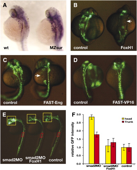
Fig. 6 FoxH1 modulates vascular formation in zebrafish. (A) flk1 is expressed in the developing vasculature of embryos at 32 hpf (left). The expression level of flk1 is elevated in MZsur embryos (right). (B) Injection of FoxH1 mRNA disrupts zebrafish vascular formation. Embryos were analyzed at the 18-somite stage. (C) Injection of mRNA encoding the FAST-Eng chimeric protein disrupts vascular formation (right), resembling the phenotype of FoxH1 overexpression. Embryos were analyzed at the 23-somite stage. (D) Injection of mRNA encoding FAST-VP16 chimeric protein does not disrupt the GFP expression pattern in TG(flk1:GFP)la116 embryos. Embryos were analyzed at the 18-somite stage. Arrows point to patches of endothelial cells missing in FoxH1 or FAST-Eng mRNA-injected embryo. (E) Lateral view of the un-injected (right), smad2MO and FoxH1 mRNA co-injected (middle) and smad2MO-injected (left) TG(flk1:GFP)la116 embryos at 24 hpf. (F) Graph represents relative GFP intensities within the region of interests (indicated by the yellow and red boxes in panel E).
Reprinted from Developmental Biology, 304(2), Choi, J., Dong, L., Ahn, J., Dao, D., Hammerschmidt, M., and Chen, J.N., FoxH1 negatively modulates flk1 gene expression and vascular formation in zebrafish, 735-744, Copyright (2007) with permission from Elsevier. Full text @ Dev. Biol.

