
Fig. 7
- ID
- ZDB-IMAGE-070219-23
- Genes
- Publication
- Nair et al., 2007 - Requirements for Endothelin type-A receptors and Endothelin-1 signaling in the facial ectoderm for the patterning of skeletogenic neural crest cells in zebrafish
- All Figures
- Figures for Nair et al., 2007
|
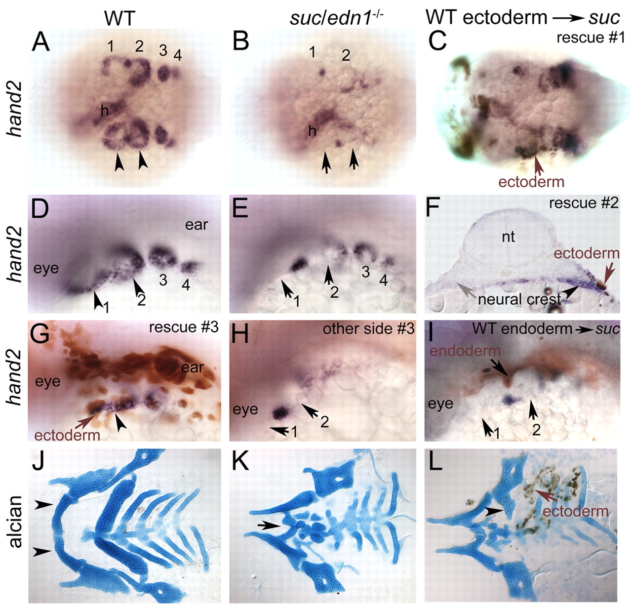
Fig. 7 Facial ectoderm is a crucial functional source of Edn1 in the arches. Whole-mount RNA in situ hybridizations at 30 hpf (A,B,D,E), with immunohistochemistry for biotin-dextran (brown cells in C,F-I). Dorsal views (A-C), dorsolateral views (D,E,G-I), 96 hpf flat-mounted Alcian-Blue-stained cartilages (J,K) combined with immunohistochemistry for biotin-dextran (L). (A,D) hand2 expression in ventral cranial NC cells in wild type (arrowheads), which is lost in suc;edn1-/- (B,E, arrows), except in few cells at arch borders. (C) Rescue of hand2 in suc;edn1-/- by unilateral grafting of ectoderm on the left side (arrow). (F) Transverse cryosection through the arch shows unilateral rescue of hand2 (arrowhead) in a suc;edn1-/- embryo adjacent to grafted ectoderm (brown cells, arrow). The control side did not receive any donor ectoderm and shows no rescue of hand2 (gray arrow). (G) Another example of wild-type ectoderm (brown cells, arrow) rescuing hand2 (arrowheads). (H) Control side of embryo in G. (I) Wild-type endoderm (brown cells, arrow) did not rescue hand2. (J) Wild-type cartilages include the mandibular, hyoid and branchial elements including Meckel's cartilage (arrowhead). (K) Meckel's cartilage is reduced in suc;edn1-/- (arrow). (L) suc;edn1-/- mutant that received wild-type ectoderm shows unilateral rescue of the ventral hyosymplectic cartilage (arrowhead) adjacent to the grafted ectoderm (arrow, brown cells). h, heart; nt, neural tube.