
Image
|
Figure Caption
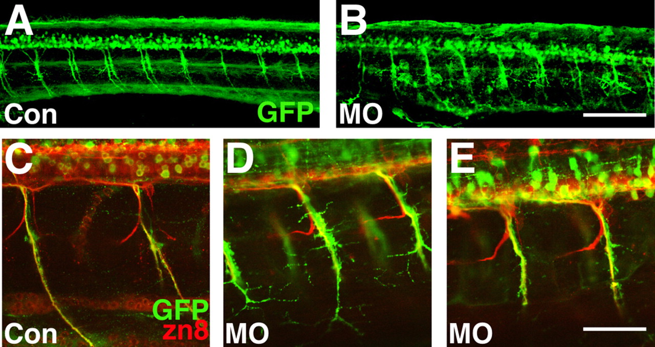
Fig. 7 Injection of 1.6MO but not ConMO perturbed outgrowth of SMN axons in Tg(gata2:GFP) embryos. (A-E) GFP+ ventrally projecting axons displayed more branching in Tg(gata2:GFP) morphant (B,D,E) but not control (A,C) embryos at 72 hpf. Embryos were squash-mounted and, consequently, motor nerves on both sides of an embryo were often present in a single confocal section (e.g. A,B). Scale bars: in B, 100 μm for A,B; in E, 50 μm for C-E.
Figure Data
Acknowledgments
This image is the copyrighted work of the attributed author or publisher, and
ZFIN has permission only to display this image to its users.
Additional permissions should be obtained from the applicable author or publisher of the image.
Full text @ Development