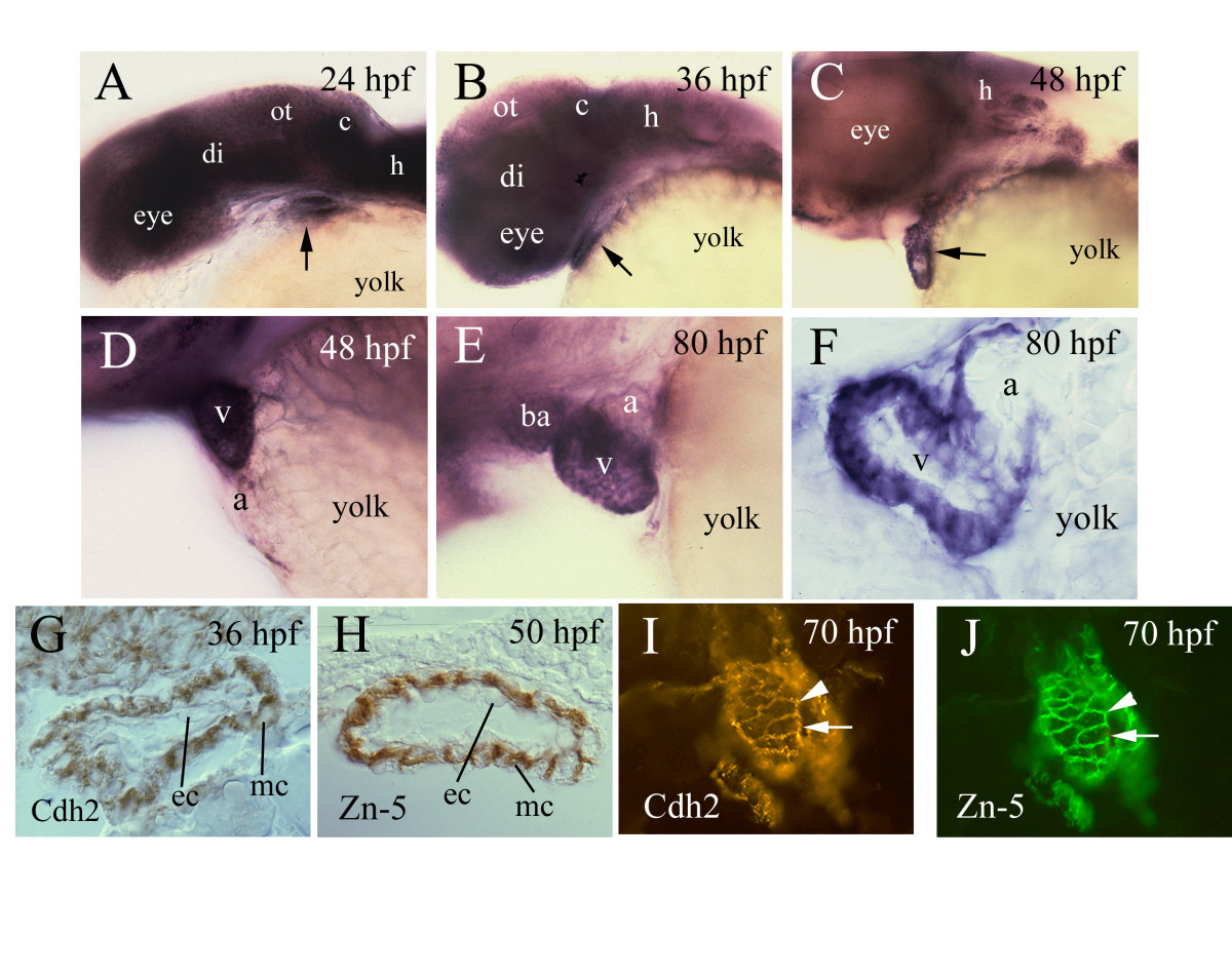
Fig. 1 Cadherin2 expression in developing zebrafish heart. Anterior is to the left and dorsal is up for panels A-H. Panels A-C are lateral views of the head region of whole mount zebrafish embryos labeled by in situ hybridization with cadherin-2 cRNA. The arrow points to the heart. Panels D and E are lateral views of higher magnifications of the heart. Panel F is a parasagittal section of a heart processed for whole mount in situ hybridization. Panels G and H are parasagittal sections of the ventricle processed for cadherin-2 (Cdh2) and Zn-5 immunocytochemical staining, respectively, both showing that the staining is confined mainly to cell membranes of myocardiocytes. Panels I and J show the same cross section of the ventricle (dorsal up) double-labeled with cadherin-2 antibody (panel I) and Zn-5 antibody (panel J). The arrows and arrowheads point to the same cells respectively. Abbreviations: a, atrium; ba, bulbus arteriosus; c, cerebellum; di, diencephalon; ec, endothelium; h, hindbrain; mc, myocardium; ot, optic tectum; v, ventricle.
Image
Figure Caption
Figure Data
Acknowledgments
This image is the copyrighted work of the attributed author or publisher, and
ZFIN has permission only to display this image to its users.
Additional permissions should be obtained from the applicable author or publisher of the image.
Full text @ BMC Dev. Biol.

