
Fig. 2
- ID
- ZDB-IMAGE-060726-4
- Publication
- Brand et al., 1996 - Mutations in zebrafish genes affecting the formation of the boundary between midbrain and hindbrain
- All Figures
- Figures for Brand et al., 1996
|
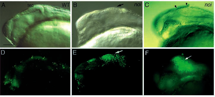
Fig. 2 Apoptosis in the tectum of noi mutant embryos. Shown is a comparison of Nomarski (A-C) and fluourescence (D-F) images of the same embryos, stained with acridine orange to detect apoptotic cell death. (A,D) Wild-type embryo at 22 h of development; (B,E) homozygous mutant noitu29a embryo at 22 h, stained with acridine orange. (C,F) Homozygous mutant noity22b embryo at 26 h, stained with acridine orange. Large numbers of stained cells are observed in the tectum of mutant embryos in E and F (arrows) that are absent in the wild type. Degenerating, more refractile cells are also seen with Nomarski optics in C (between arrowheads). Scale bar, 120 µm.