
Fig. 4
- ID
- ZDB-IMAGE-060628-27
- Genes
- Antibodies
- Publication
- Wei et al., 2004 - The zebrafish Pard3 ortholog is required for separation of the eye fields and retinal lamination
- All Figures
- Figures for Wei et al., 2004
|
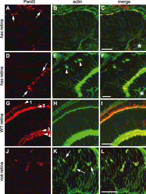
Fig. 4 Pard3 immunolocalization in has and nok mutant retinas. Immunohistochemistry was performed on 33 hpf (A–C) and 4 dpf (D–F) hasm567 mutant retinas, 4 dpf wild-type retinas (G–I), and 33 hpf nokm520 mutant retinas (J–L) with anti-Pard3 antiserum (left column) and Alexa-488-conjugated phalloidin (middle column). The merged images are shown in the right column. Pard3 (A, arrows) localized to the apical region in the hasm567 retinal neuroepithelium at 33 hpf and partially overlapped with adherens junction-associated actin bundles. At 4 dpf, Pard3 and actin were both localized in the inner plexiform layer in the hasm567 retina (D, arrows), while actin staining in the outer plexiform layer was interrupted (E, arrowheads). In the 4 dpf wild-type retina, Pard3 and actin localized to the outer limiting membrane (arrowhead 1), the outer plexiform layer (arrowhead 2), and two sublayers in the inner plexiform layer (arrowheads 3 and 4). In the nokm520 mutant retinal neuroepithelium, Pard3 localized to the ectopic adherens junction-associated actin bundles (K, arrows). Scale bars indicate 20 μm.
Reprinted from Developmental Biology, 269(1), Wei, X., Cheng, Y., Luo, Y., Shi, X., Nelson, S., and Hyde, D.R., The zebrafish Pard3 ortholog is required for separation of the eye fields and retinal lamination, 286-301, Copyright (2004) with permission from Elsevier. Full text @ Dev. Biol.