
Fig. 4
- ID
- ZDB-IMAGE-060620-3
- Publication
- Villablanca et al., 2006 - Control of cell migration in the zebrafish lateral line: Implication of the gene "Tumour-Associated Calcium Signal Transducer," tacstd
- All Figures
- Figures for Villablanca et al., 2006
|
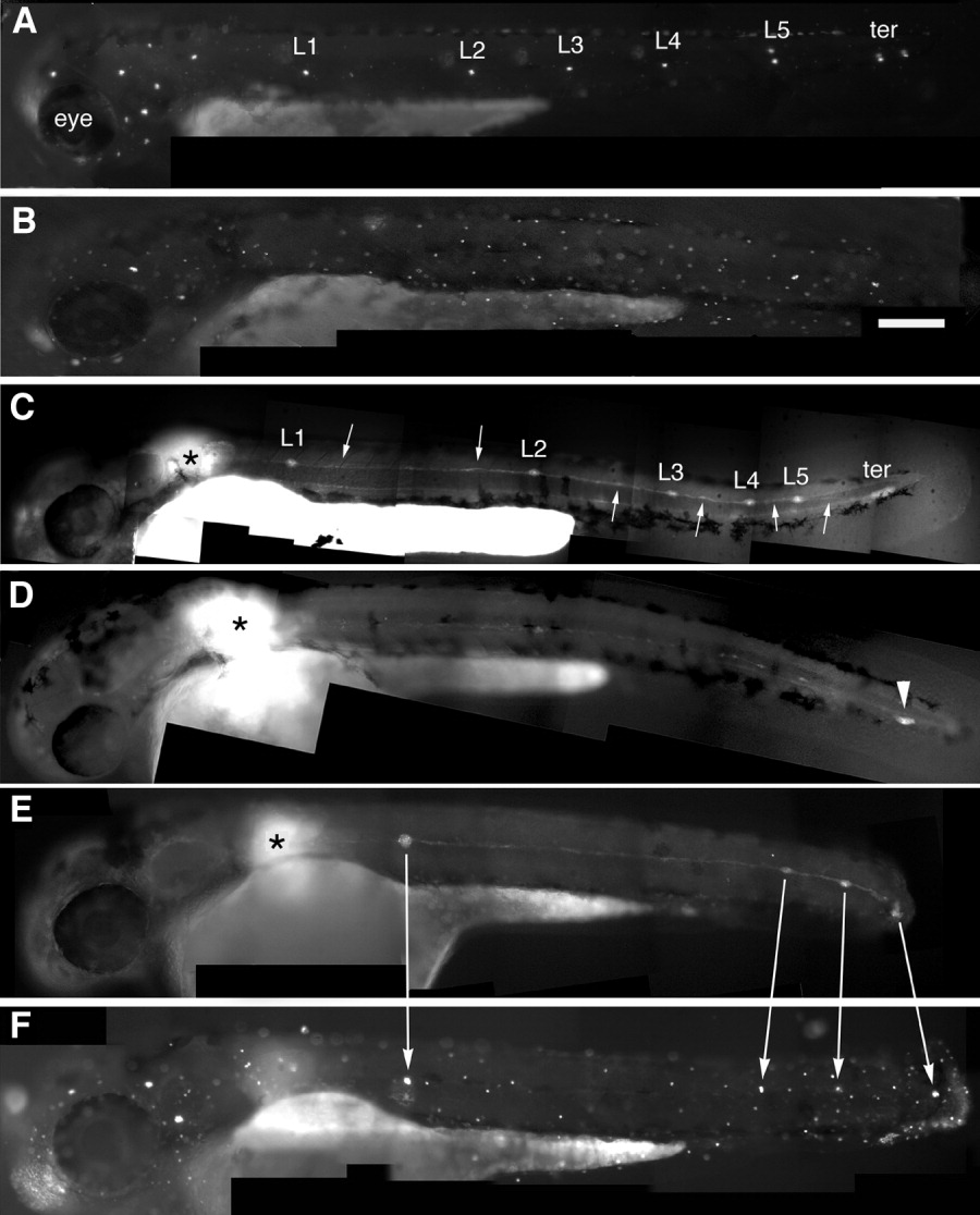
Fig. 4 Embryonic posterior lateral line labeled by 4-Di-2-ASP, a marker of differentiated hair cells (A, B, and F) or by uncaging the primordium (C-E). A: Wild type embryo with the normal pattern of five neuromasts along the horizontal myoseptum (L1-L5) and two terminal neuromasts (ter). B: Morphant embryo where all PLL neuromasts are missing. C: Uncaging the primordium at the position marked by the asterisk in a wild type embryo reveals the neuromasts (L1-5 and ter) and interneuromastic cells (arrows). D: Uncaging in a morphant reveals that the primordium has reached the tip of the tail (arrowhead) but no neuromasts have been deposited. E,F: In a partial morphant where four clusters have been deposited, including a terminal one (E), each cluster has differentiated hair cells on the next day (F).