
|
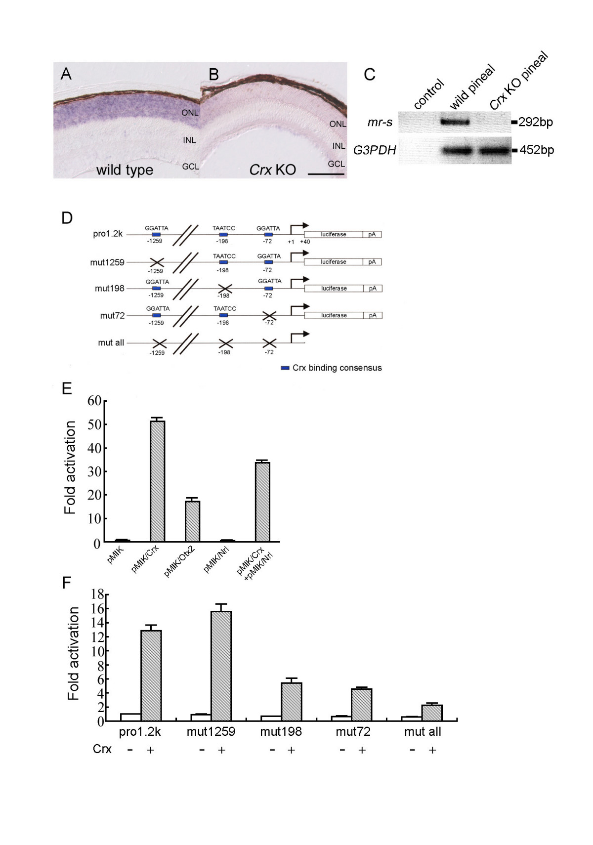
Fig. 3 The transcription of mr-s is regulated by Crx. (A, B) In situ hybridization using a probe for mouse mr-s was performed on the wild-type (A) and Crx KO retinas (B) at P5. mr-s expression was drastically reduced in the Crx KO retina (B). Scale bar, 100 μm. (C) RT-PCR analysis of total RNAs extracted from the pineal glands of P5 wild-type and Crx KO mouse. The upper and lower lanes show PCR products amplified by the primer pairs specific for mr-s and G3PDH cDNAs, respectively. Water was used for control RT-PCR reaction. (D-F) Crx transactivates mr-s transcription. Reporter plasmids for the luciferase assay are shown. Blue boxes represent Crx binding sites (D). Relative activity of the luciferase is indicated when Pro1.2k was co-transfected with Crx, Otx2, Nrl, and Crx+Nrl, respectively (E). Fold activation is indicated when Pro1.2k, mut1259, mut198, mut72 and mut all were co-transfected with the Crx expression vector (Crx+) or the empty vector (Crx-) into HEK293T cells (F). Error bars represent standard error of mean.