
Fig. 3
- ID
- ZDB-IMAGE-060424-14
- Genes
- Publication
- Grinblat et al., 1998 - Determination of the zebrafish forebrain: induction and patterning
- All Figures
- Figures for Grinblat et al., 1998
|
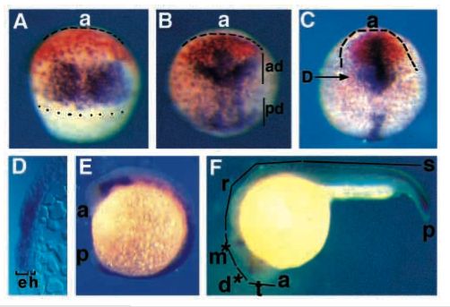
Fig. 3 opl and fkh5 are expressed in adjacent domains that mark presumptive telencephalon and diencephalon at gastrula. Staged embryos were stained for opl RNA (orange) and fkh5 RNA (purple) using whole-mount in situ hybridization. (A) Mid-gastrula (75% epiboly) embryo, dorsal view, dotted line marks blastoderm margin; dashed line outlines anterior neural plate. (B) Late gastrula (90% epiboly) embryo, dorsal view; ad, anterior domain of fkh5 expression; pd, posterior domain of fkh5 expression. (C) Early neurula (tailbud) embryo, dorsal view, arrow shows plane of section shown in D. (D) Transverse section through a tailbud stage embryo. Note that staining is strong in the epiblast (e) and excluded from hypoblast (h). (E) Early somitogenesis (5 somite) embryo, side view. (F) Late somitogenesis (prim-5) embryo, side view, asterisks indicate weak fkh5 staining. a, anterior; t, telencephalon; d, diencephalon; m, mesencephalon; p, posterior; r, rhombencephalon; s, spinal cord.