
Fig. 7
- ID
- ZDB-IMAGE-060410-29
- Genes
- Publication
- Takamiya et al., 2006 - Hedgehog signalling controls zebrafish neural keel morphogenesis via its level-dependent effects on neurogenesis
- All Figures
- Figures for Takamiya et al., 2006
|
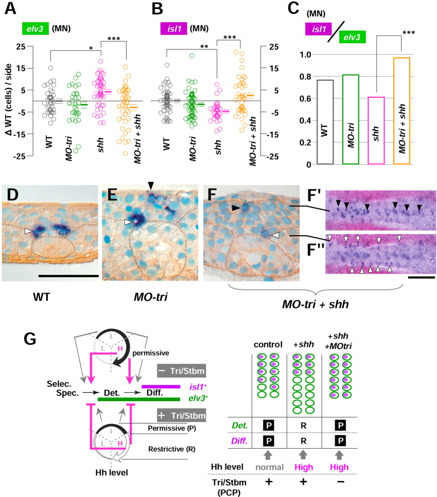
Fig. 7 Medial Tri/Stbm mediated planar cell polarity (PCP) pathway reverses hedgehog (Hh) function on the medial neurogenesis into opposite in a polarity-independent manner. A-C: The number of neurons (either elv3-positive or isl1-positive) are counted during the neural keel stage (4- to 10-somite stages) and analyzed in mediolateral distance (MLD) time. As in Figures 5 and 6, results are shown as the difference from the uninjected controls of the identical MLD stages ({delta}WT [wild-type], cells/side) and pooled for each group of embryos of WT, MO-tri, shh mRNA-injected, and MO-tri/shh embryos. P values from statistical analysis by Tukey′s multiple comparison test after one-way analysis of variance (A,B) are shown with symbols: ***, P < 0.001; **, P < 0.01; and *, P < 0.05. Note that in shh mRNA-injected embryos, the increase from uninjected controls for elv3 (A, *P < 0.05) and decrease in isl1 cells (B, **P < 0.01) turned into the opposite upon co-injection with MO-tri, which are clearly reflected in motoneuron (MN) differentiation index (ratio of isl1 medial cells to elv3 medial neurons, C). D-F: Ectopic medial neurons are repressed by Tri/Stbm-mediated PCP pathway in a polarity-independent manner. D: In uninjected controls, medial isl1-PMNs are found (white arrowhead) at the bottom (basal side) of the neural keel intervened by floor plate cells adjacent to the notochord (dotted circle) and usually never seen at the apical side. E: However, in MO-tri embryos, ectopic isl1-medial neurons (black arrowhead) are found at the apical surface, together with the normal PMN at the basal position (white arrowhead). F,F′: This effect seems to be by means of a polarity-independent pathway, because ectopic medial neurons were not suppressed (black arrowhead; dorsal views at the focal plane of the line) in embryos with disturbed cell polarity (MO-tri/shh embryos) to a level comparable to shh embryos with no ectopic medial neurons. G: (Left) Medial Tri/Stbm-mediated PCP pathway reverses the function of a high Hh level at the two steps of medial neurogenesis: the establishment of commitment (at the transition from specification to determination) and the onset of neuronal differentiation (at the transition from determination to differentiation). As to the function of a low Hh level, analysis is pending (indicated by dotted line). (Right) Schematic drawings to present how reversed Hh function upon reduced Tri/Stbm-mediated PCP pathway caused supernumerary medial isl1 neurons. elv3 expression is shown as blank green ellipses and isl1 expression as violet-filled ellipses. P, permissive; R, restrictive. Scale bars = 50 μm for D-F, 100 μm for F.