
Fig. 8
- ID
- ZDB-IMAGE-060410-16
- Genes
- Publication
- Hammond et al., 2006 - The developing lamprey ear closely resembles the zebrafish otic vesicle: otx1 expression can account for all major patterning differences
- All Figures
- Figures for Hammond et al., 2006
|
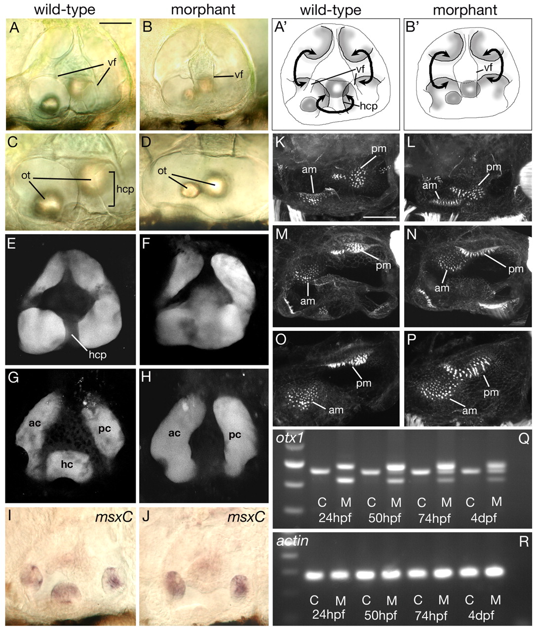
Fig. 8 Effects on the developing ear of injection of otx1 morpholino into wild-type zebrafish embryos. (A-D) DIC images of ears of live embryos. (A',B') Schematic representation of ears shown in A,B. Note the lack of the horizontal canal projection (hcp) that delimits the horizontal semicircular canal and the fusion of the two ventral folds (vf) in morphant embryos. The otoliths (ot) of morphants are also closer together than in wild type. Curved black arrows indicate the semicircular canal lumens. (E-H) Confocal sections through ears in which the lumens have been filled with fluorescein-conjugated morpholino. (E,F) Medial sections, in the plane of the semicircular canal projections; (G,H) Lateral sections, in the plane of the lumens of the semicircular canals. ac, anterior canal; pc, posterior canal; hc horizontal canal. (I,J) mRNA expression pattern of msxC marking the cristae. (K-P) Projections of confocal z-stacks of FITC-phalloidin-stained ears revealing the actin-rich stereocilia of the sensory hair bundles. am, anterior macula; pm, posterior macula. (A-L) Lateral views, dorsal to top; (M,N) dorsal views, medial to top; (O,P) junction between anterior and posterior maculae. Anterior is to the left in all panels. All ears are at 96 hpf, except I and J, which are 72 hpf. Scale bars: 50 μm. (Q) RT-PCR across the exon 3/4 boundary. Lane 1, 100 bp ladder; brightest band is 500 bp. (R) RT-PCR using primers to actin. C, control uninjected siblings; M, morpholino-injected embryos.