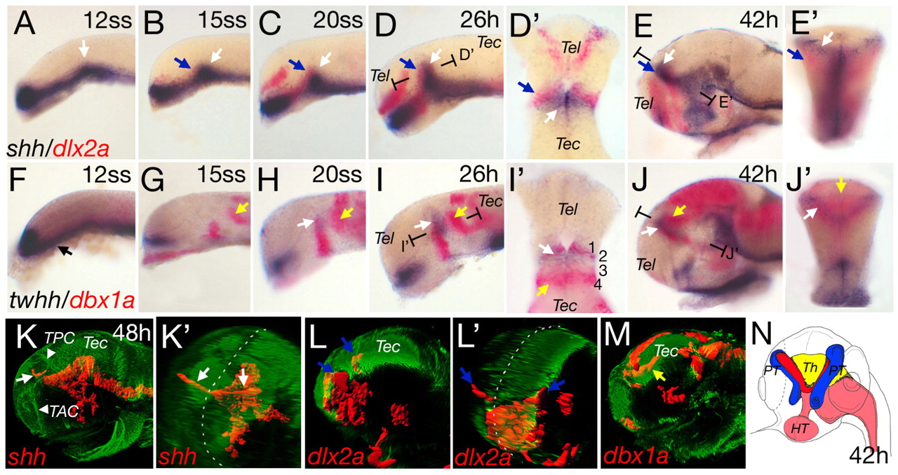
Fig. 1 Anteroposterior differentiation in the mid-diencephalic territory (MDT). Whole-mount double in situ hybridization of wild-type embryos, with shh and dlx2a (A-E′) and twhh and dbx1a (F-J′). Section planes of D′,E′,I′,J′ are indicated. (A) shh expression in the ventral neural tube with the position of the presumptive ZLI indicated (white arrow). At the 15-somite stage, dlx2a expression starts in the forebrain (B,C; blue arrows). Horizontal section of D shows the abutting expression domains of dlx2a and shh (D′). shh expression extends to the dorsal and dlx2a expression is located more laterally (E, blue arrow). A cross-section in (E′) shows the ZLI (white arrow) and the prethalami (blue arrow). Onset of twhh expression in the presumptive ZLI is contemporaneous with shh (H and I, white arrow). twhh expression marks the v-shaped structure of the ZLI at 42hpf (J,J′). At 12 somites, dbx1a expression is anteroventral (F,G). At 15 somites, dbx1a is expressed at the base of the future ZLI (G), extending dorsally in later stages (H-J) and co-localized with twhh [horizontal section at 26 hpf (I′) and cross-section at 42 hpf (J′)]. From 15 somites onwards, a posterior domain of dbx1a can be detected, marking the presumptive thalamus (G-J, yellow arrows). At 42 hpf, the dbx1a-positive thalamus lies in a medial position, posterior to the ZLI (cross-section in J′). (K-M) Lateral and pseudo-frontal views of 3D reconstructions of confocal stacks of in situ hybridization combined with anti-acetylated tubulin staining. Broken white lines indicate the midline (K′ and L′). At 48 hpf, shh is expressed in the ZLI in two prongs (K,K′, white arrows). dlx2a expression in the prethalami is located ventral and lateral to the ZLI (L,L′, blue arrows). dbx1a is expressed posterior and dorsal to the shh expression domain (M, yellow arrow). (N) Scheme of the subdomains of the MDT based on marker expression at 42 hours: red, ZLI; blue, prethalami; yellow, thalamus; pink, shh-positive basal plate. HT, hypothalamus; PT, prethalamus; Th, thalamus; TAC, tract of the anterior commissure; Tec, tectum; Tel, telencephalon; TPC, tract of the posterior commissure. Embryos are oriented dorsal side to the top and anterior towards the left in all figures, except when indicated.
Image
Figure Caption
Figure Data
Acknowledgments
This image is the copyrighted work of the attributed author or publisher, and
ZFIN has permission only to display this image to its users.
Additional permissions should be obtained from the applicable author or publisher of the image.
Full text @ Development

