
Image
|
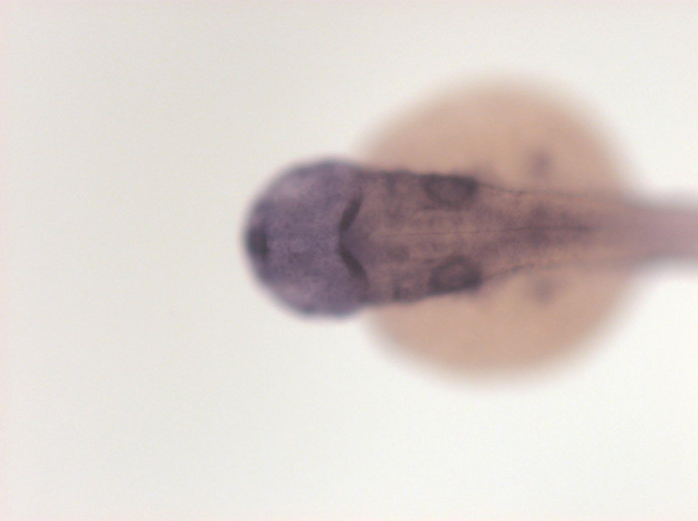
Figure Caption
Fig. 5 caudal most notochord, spinal chord, branchial arches, lateral line primordia, otic vesicle, dorsal diencephalon and midbrain, optic stalks, hypothalamus, nuclei in diencephalon, pharyngeal pouches
Orientation
| Preparation | Image Form | View | Direction |
| whole-mount | still | dorsal | anterior to left |
Figure Data