
Fig. 3
- ID
- ZDB-IMAGE-050930-9
- Genes
- Antibodies
- Publication
- Babb et al., 2005 - Zebrafish R-cadherin (Cdh4) controls visual system development and differentiation
- All Figures
- Figures for Babb et al., 2005
|
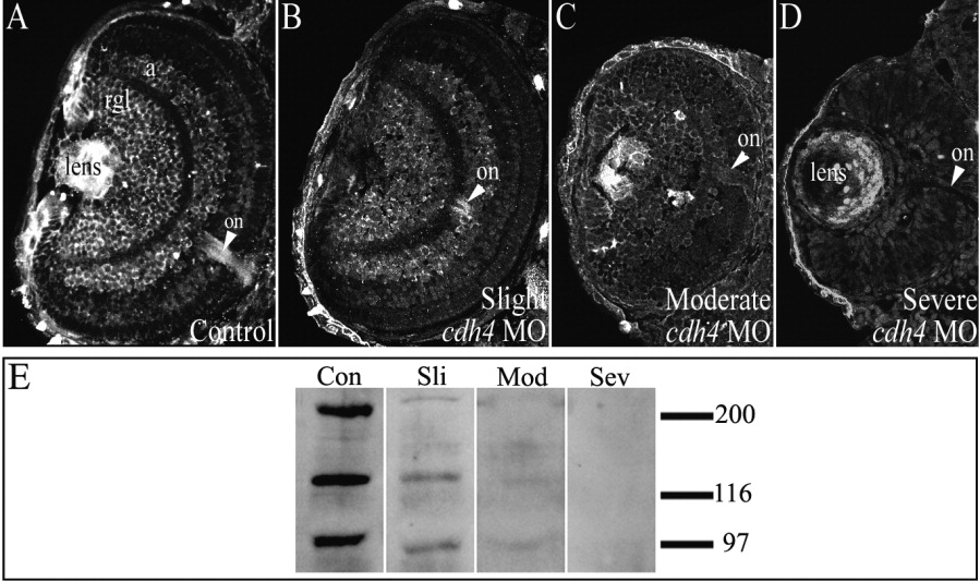
Fig. 3 Reduction of Cdh4 protein levels correlates with phenotypic severity of cdh4 morphants. A-D: Zebrafish embryos were injected with either standard control morpholino oligonucleotide (MO, A) or cdh4 MO (B-D). A: Confocal imaging of 3 days postfertilization (dpf) Cdh4 immunostained cryosections showed Cdh4 localization in the retinal ganglion cell layer, in the amacrine cell layer of the inner nuclear layer, in the lens, and in the optic nerve of the control embryo. B,C: In slightly and moderately affected cdh4 MO-injected embryos, staining of the retinal ganglion cell layer, the amacrine cell layer and the optic nerve was reduced. D: In severely affected embryos, Cdh4 staining was not detected in the neural retina and reduced in the lens (the apparent immunoreactivity of the ectoderm and lens is nonspecific, that is, it could not be competed with excess immunizing peptide; Liu et al.,[2001b]). E: Corresponding immunoblots of individual embryos of various phenotypic severities in the hypomorphic series showed that, compared with control embryos at 3 dpf (con), Cdh4 expression was reduced in slightly and moderately affected embryos (sli, mod) and absent in severely affected embryos (sev).