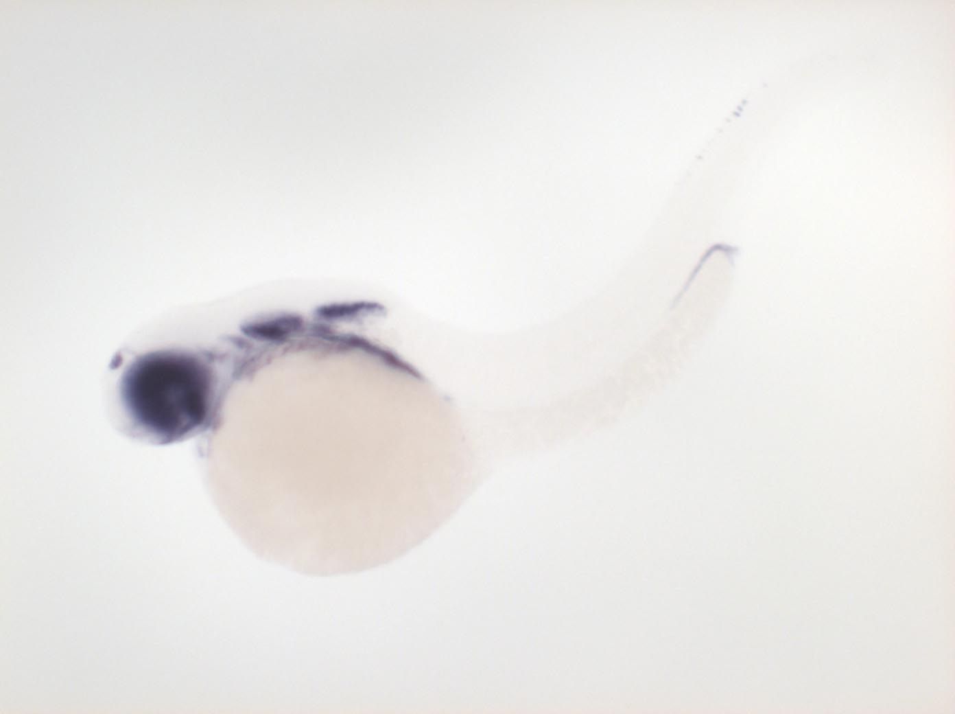
Image


                        Figure Caption


Fig. 5 Expressed in ventral anterior diencephalon, posterior retina, epiphysis, otic vesicle, part of pharyngeal arches 3-7, pectoral fin bud, dorsal spinal cord neurons, posterior pronephric ducts, anterior spinal cord, head muscles
                
                    
                        Orientation


                | Preparation | Image Form | View | Direction | 
| whole-mount | still | side view | anterior to left | 
                
                    
                        Figure Data


        