Image
Figure Caption
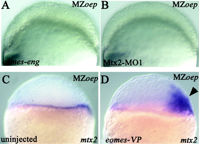
Fig. 5 Epiboly defect is Nodal-independent. All views are lateral; the mutant phenotype is indicated in upper right corner; the injected construct is indicated in lower left corner. A,B: At 50% epiboly (5.25 hours postfertilization [hpf]). C,D: At sphere stage (4 hpf). A: Embryo injected with eomes-eng; the blastoderm has failed to thin. B: Embryo injected with Mtx2-MO1; the blastoderm has failed to thin. C: mtx2 expression in an uninjected embryo. D: Ectopic mtx2 expression (arrowhead) in an eomes-VP-injected (into a single cell at the eight-cell stage) embryo.
Figure Data
Acknowledgments
This image is the copyrighted work of the attributed author or publisher, and
ZFIN has permission only to display this image to its users.
Additional permissions should be obtained from the applicable author or publisher of the image.
Full text @ Dev. Dyn.

