
Fig. 4
- ID
- ZDB-IMAGE-050323-18
- Publication
- Waxman et al., 2004 - Zebrafish Dapper1 and Dapper2 play distinct roles in Wnt-mediated developmental processes
- All Figures
- Figures for Waxman et al., 2004
|
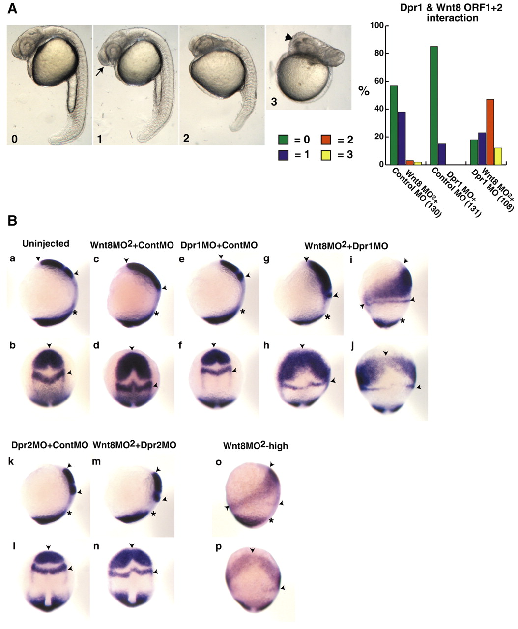
Fig. 4 Dpr1 is an enhancer of Wnt/ß-catenin signaling required for ventral and posterior cell fates. (A) Representative embryos for scoring of ventroposteriorization at 24 hours. 0, wild type; 1, slight enlargement of the telencephalon (arrow); 2, strong anteriorization phenotype, enlargement of the head and reduction of the tail; and 3, very strong anteriorization phenotype, super enlargement of the telencephalon (arrowhead) and major loss of trunk and tail. Graph shows percentage of embryos with the respective phenotypes (numbers in brackets indicate the number of embryos used). (B) dpr1, dpr2 and wnt8MO2 morphant phenotypes at the one- to two-somite stage. In situ markers used were opl (telencephalon), pax2.1 (midbrain/hindbrain boundary) and tbx6 (ventrolateral mesoderm). Arrowheads indicate distance between anterior limit of opl and posterior limit of pax2.1. Asterisks indicate anterior limit of tbx6. (a,c,e,g,i,k,m,o) Dorsal is towards the right. (b,d,f,h,j,l,n,p) Dorsal is out of the plane of the page. In all figures, anterior is upwards. Suboptimal dose of wnt8 MO2 is 0.45 ng each MO (0.9 ng total). dpr1 MO dose is 12 ng. dpr2 MO dose is 8 ng. The experiments were repeated five times with comparable results; see text for penetrance of phenotypes.