
|
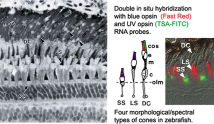
Fig. 1 Spectral subtypes of photoreceptors in zebrafish. Radial, histological section of adult zebrafish retina, with the retinal pigmented epithelium and choroid at the top. Rod nuclei (r) in the outer nuclear layer, are connected by thin myoid processes to ellipsoids (arrowheads) and rod outer segments (ros). The four morphological/spectral cone subtypes are shown in the cartoon, with the cone outer segments (cos) color-coded to indicate the spectral peak of the opsin protein expressed in short single (SS), long single (LS) and double cones (DC). Other abbreviations: ellipsoid, e; myoid, m; cone nucleus, c; outer limiting membrane (olm) of the retina. The insert shows double in situ hybridization with two different opsin RNA probes and fluorescent detection. Methods: RNA probes are tagged with digoxigenin (DIG) or fluorescein (FL), respectively, and are detected with anti-DIG and anti-FL antibodies conjugated to horseradish peroxidase (HRP) or alkaline phosphatase (AP), respectively, and visualized with enzymatic substrates that produce contrasting Fig. 2. Cone mosaic pattern in zebrafish. En face views of isolated, flattened, adult zebrafish retinas, processed for in situ hybridization with cone opsin RNA probes (fluorescent signals are pseudo-colored to correspond to the spectral subtype). At lower left is a cartoon schematic of the cone mosaic pattern. fluorescent precipitates (Barthel and Raymond, 2000; Jowett, 2001). The Fast Red AP substrate is bright red in visible light, and it also fluoresces red with a rhodamine filter set. The TSA Biotin System uses HRP to catalyze the deposition of biotin-labeled tyramide, which is recognized by a streptavidin-fluorophore conjugate, such as fluorescein isothiocyanate (FITC). We performed control experiments to verify that the different detection methods have comparable sensitivity.