
|
OPTICS: MAGNIFICATION: DATE OF IMAGE: SUBMITTER COMMENTS:
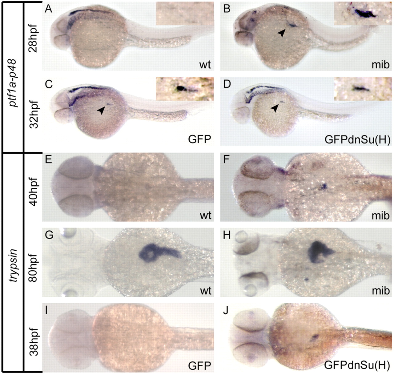
Fig. 6 Defects in Notch pathway activation result in acceleration of exocrine differentiation in developing zebrafish pancreas. (A-D) Mindbomb (mib) mutant embryos (B) and embryos injected with RNA encoding a dominant-negative Suppressor of Hairless DNA binding mutant [GFPdnSu(H)] (D) show either accelerated (mib) or normal [GFPdnSu(H)] onset of ptf1a-p48 expression compared with clutchmate controls (A,C). Arrows indicate endodermal domain of ptf1a-p48 expression, distinct from expression in developing hindbrain. Insets in A-D show magnified view of endodermal ptf1a-p48 expression domain. (E-J) mindbomb (mib) mutant embryos (F,H) and embryos expressing GFPdnSu(H) (J) show accelerated acinar cell differentiation compared with clutchmate controls (E,G,I), marked by early onset of trypsin expression. Note normal absence of trypsin expression in control embryos at 40 hpf (E,I), but accelerated onset of trypsin expression in mibta52/ta52 and GFPdnSu(H)-injected embryos (F,J). At 80 hpf, the size and contour of established trypsin-positive exocrine parenchyma is also altered in mibta52/ta52 embryos compared with wild-type clutchmates (G,H). Wt indicates wild-type clutchmates arising from mibta52/wt x mibta52/wt cross. GFP indicates clutchmate control embryos injected with RNA encoding GFP alone.
| Preparation | Image Form | View | Direction |
| not specified | still | not specified | not specified |