
Figure 5
- ID
- ZDB-FIG-260623-14
- Publication
- Capecki et al., 2026 - Evidence that disruption of Discoidin domain receptor 2 contributes to palate malformations through effects on the extracellular matrix
- Other Figures
- All Figure Page
- Back to All Figure Page
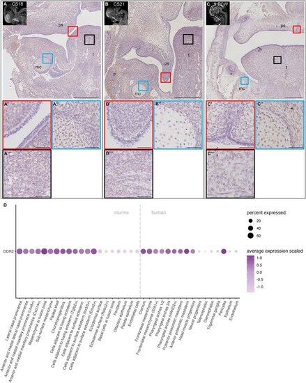
DDR2 expression analysis in human and murine embryos. (A–C) RNA-scope-in-situ (brown staining) of cranial human embryo sections at Carnegie-stage (CS) 18 and 21, as well as 9 weeks post conception (PCW). The approximate plain of sections is marked with a white line in the upper left corner. For each image, magnified palatal shelf (ps; A’, B′, C′), Meckel’s (mc; A”, B″, C″), and tongue (t; A”’, B”’, C”’) areas are shown. Scale bars: Overviews, 500 μm; magnified regions, 50 μm. (D) Dotplot of DDR2 gene expression in the lambdoid junction of embryonic day 11.5 murine embryos and in the head of CS12–16 human embryos. The size of the dots corresponds to the percentage of cells expressing the gene in the respective cell type. Dot color intensity represents the average scaled expression level. |