
Fig. 5
- ID
- ZDB-FIG-260602-18
- Publication
- Zhao et al., 2026 - Atf4a Regulates Mitochondrial Homeostasis Through Parkin-Mediated Mitophagy to Enhance Hypoxia Tolerance in Zebrafish (Danio rerio)
- Other Figures
- All Figure Page
- Back to All Figure Page
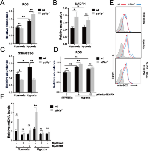
Atf4a deficiency leads to severe mitochondrial oxidative stress in zebrafish. (A) Mitochondrial reactive oxygen species (ROS) levels, measured by MitoSOX staining, in wt and atf4a−/− embryos treated with normoxia or hypoxia at 72 hpf. (B) Relative mean value of NADPH expression levels in wt and atf4a−/− treated normoxia and hypoxia at 72 hpf. (C) Relative GSH/GSSG ratio in wt and atf4a−/− treated normoxia and hypoxia at 72 hpf. (D-E) Mitochondrial ROS levels, measured by MitoSOX staining and flow cytometry, in wt and atf4a−/− zebrafish embryos, with or without 100 μM mito-TEMPO supplementation at 72 hpf. Quantification of mitochondrial ROS levels is shown in (D), and representitive flow cytometry histograms are shown in (E). Gray shading indicates unstained cells. (F) Relative mitochondrial DNA (mtDNA) levels in wt and atf4a−/− zebrafish embryos treated with normoxia or hypoxia, with or without 10 μM N-acetylcysteine (NAC) or 10 μM butylated hydroxytoluene (BHT) supplementation, at 72 hpf. Data represent the mean ± SEM of three independent experiments. Unpaired two-tailed Student's t-test was used for statistical analysis. ns, not significant. *p < 0.05, **p < 0.01 indicate a significant difference between the indicated groups. |