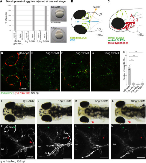
Injection of T-DM1 interrupts the development of zygotes and ablates BLECs in zebrafish embryos. (A) Injection of T-DM1 into zygotes leads to a halt of embryonic development at the one-cell stage, as cell division is suppressed [IgG-A647, n=179 (100%) zygotes with normal development; 2 ng T-DM1, n=129 (100%) zygotes with halt in development; 0.2 ng T-DM1, n=5 (2,67%) zygotes with normal development and n=182 zygotes (97.32%) with halt in development]. (B) Schematic representation of a zebrafish embryo (dorsal view) showing the injection site into the CSF and the arrangement of dorsal BLECs distal to the TeO. (C) Schematic lateral view of a zebrafish embryo depicting BLECs on the dorsal (light green) and ventral (dark green) side of the brain and facial lymphatics in red. (D-G) Maximum projections of fli:nucGFP; lyve1:dsRed-positive embryos at 5 dpf that were injected with IgG-Alexa-647 (D), or with 1 ng (E), 5 ng (F) or 10 ng (G) T-DM1 at 3 dpf. (H) The injection of T-DM1 leads to a significant reduction in the number of dorsal BLECs. n=10 embryos per group. IgG-Alexa-647 versus 1 ng T-DM1 ***P.adj=0.000966; IgG-Alexa-647 versus 5 ng T-DM1 ***P.adj=0.00087; IgG-Alexa-647 versus 10 ng T-DM1 ***P.adj=0.0009 (Mann–Whitney U-test; data are mean±s.d.). (I-L′) Bright-field images (dorsal views, I-L) and maximum projections of lyve1:dsRed-positive embryos (lateral views, I′-L′) at 120 hpf that were injected with IgG-Alexa-647 as control (I,I′), or with 1 ng (J,J′), 5 ng (K,K′) or 10 ng T-DM1 (L,L′), showing that high concentrations of T-DM1 (5 ng and 10 ng) not only ablate BLECs (green asterisks) but also affect facial lymphatic structures (red asterisks), resulting in edema formation at 5 dpf (red arrows in bright-field images). 1 ng T-DM1 ablates BLECs but does not affect the OLV (red arrows). CSF, cerebrospinal fluid; FCLV, facial collecting lymphatic vessel; hpf, hours post-fertilization; LAA, lymphatic branchial arches; LFL, lateral facial lymphatics; MFL, medial facial lymphatic; s.d., standard deviation; TeO, tectum opticum; OLV, otolithic lymphatic vessel.
|
