
Fig. 3
- ID
- ZDB-FIG-251022-16
- Publication
- Seaver et al., 2025 - Overexpression of potassium channel Kcna5 alters skeletal patterning in the zebrafish regenerating fin
- Other Figures
- All Figure Page
- Back to All Figure Page
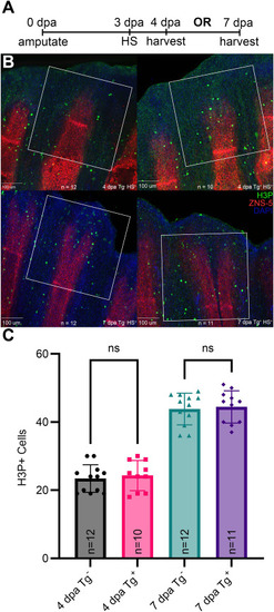
Proliferation is unchanged following overexpression of exogenous Xl-kcna5. A) Overview of experimental design. At day 0, fins from non-transgenic and transgenic siblings were amputated at the 50 % level and were systemically heat shocked at 37 °C for 1 h at 3 dpa. Fins were harvested and fixed at 4 dpa/1 dpt or at 7 dpa/4 dpt and processed for immunofluorescence. B) Fins were stained with the anti-H3P antibody to detect mitotic cells (green) and with the ZNS5 antibody to detect skeletal cells (red). C) Counts of H3P positive cells were taken from 250 μm squares from the distal tip in maximum intensity projections of confocal images. Neither timepoint showed significant differences between mitotic cells between transgenic and non-transgenic siblings. dpa, days post-amputation. (For interpretation of the references to colour in this figure legend, the reader is referred to the Web version of this article.) |
Reprinted from Developmental Biology, , Seaver, A.W., Li, X., Iovine, M.K., Overexpression of potassium channel Kcna5 alters skeletal patterning in the zebrafish regenerating fin, , Copyright (2025) with permission from Elsevier. Full text @ Dev. Biol.