
FIGURE
Fig. 1
- ID
- ZDB-FIG-251016-93
- Publication
- Bowley et al., 2021 - Quantifying endothelial cell proliferation in the zebrafish embryo
- Other Figures
- All Figure Page
- Back to All Figure Page
Fig. 1
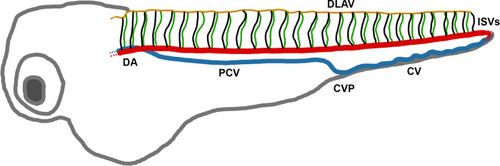
Diagram of the zebrafish vasculature at 72 hpf. DA – dorsal aorta. DLAV - dorsal longitudinal anastomotic vessel. CV - caudal vein. CVP - caudal vein plexus. PCV - posterior cardinal vein. ISVs - intersegmental vessels. |
Expression Data
Expression Detail
Antibody Labeling
Phenotype Data
Phenotype Detail
Acknowledgments
This image is the copyrighted work of the attributed author or publisher, and
ZFIN has permission only to display this image to its users.
Additional permissions should be obtained from the applicable author or publisher of the image.
Full text @ F1000Res