
Fig. 4
- ID
- ZDB-FIG-250909-60
- Publication
- Collins et al., 2025 - FBXW7 regulates MYRF levels to control myelin capacity and homeostasis in the adult central nervous system
- Other Figures
- All Figure Page
- Back to All Figure Page
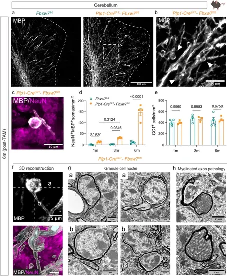
Loss of Fbxw7 results in ectopic ensheathment of neuronal cell bodies in the cerebellum. a Representative images of anti-MBP stained cerebellums from Fbxw7fl/fl and Plp1-CreERT-Fbxw7fl/fl animals at 6-months post-TAM. Dotted line denotes the border of the granule layer. b Higher magnification image of MBP staining in the granular layer of a Plp1-CreERT-Fbxw7fl/fl animal showing cupped myelin structures, which surround NeuN+ nuclei (shown at higher magnification in c). d Quantification of NeuN+ cells wrapped in MBP positive membrane in the granular layer of the cerebellum of Fbxw7fl/fl (1 m: N = 4, 3 m: N = 3, 6 m: N = 4) and Plp1-CreERT-Fbxw7fl/fl mice (1 m: N = 4, 3 m: N = 3, 6 m: N = 4) at 1-, 3-, and 6-months post-TAM. e Quantification of CC1+ OLs in the white matter and granular layer of Fbxw7fl/fl (1 m: N = 4, 3 m: N = 3, 6 m: N = 4) and Plp1-CreERT-Fbxw7fl/fl mice (1 m: N = 4, 3 m: N = 3, 6 m: N = 4) at 1-, 3-, and 6-months post-TAM. Data shown as average ± SEM. Statistical significance determined by two-way ANOVA. f Image of MBP+ myelin outfold (white) around a granule cell (magenta) by IF at 6-months post-TAM reconstructed in Imaris. a and b dotted lines represent orientation of EM images in g. g Representative images of myelin outfolds around granule cell nuclei in the granule layer of the cerebellum at 6-months post-TAM. This was observed in all 4 mutant mice animals analyzed. h Images of severe cellular accumulations in the axons of putative Purkinje cells in Plp1-CreERT-Fbxw7fl/fl mice at 6-months post-TAM. This was observed in all mutant animals analyzed (N = 4). Created in BioRender. Emery, B. (2024) BioRender.com/o45h010/k49b495. |