
Fig. 2
- ID
- ZDB-FIG-250605-8
- Publication
- Tang et al., 2025 - Visualization of Unhatched Brine Shrimp Eggs in Zebrafish Intestines Using Synchrotron Radiation Phase-Contrast CT
- Other Figures
- All Figure Page
- Back to All Figure Page
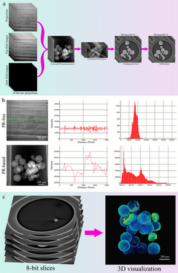
Image processing and reconstruction. (a) PR-based PCCT slices acquisition. PR processing was performed to acquire PR-based projections before the sinograms were generated. The floating-point sinograms were reconstructed into floating-point slices. Then the floating-point slices were converted to 8-bit slices. (b) Comparison of intensity variation and histogram between PR-free and PR-based projections. Line profile analyses along the green dotted lines were displayed. Note that PR processing enabled a greater intensity difference between different tissues, although the PR-free projection had more detailed intensity information. The histogram from the PR-based projection had a wider range of intensity distribution than that from the PR-free projection. (c) 3D reconstruction. 3D visualization was obtained by reconstructing the 8-bit PR-based PCCT slices. |