
FIGURE
Fig. 4
- ID
- ZDB-FIG-250422-124
- Publication
- Guo et al., 2025 - Fluorine-Nitrogen Codoped Carbon Dots for Visualization Imaging of Nucleic Acids via Two-Photon Fluorescence Lifetime Microscopy
- Other Figures
- All Figure Page
- Back to All Figure Page
Fig. 4
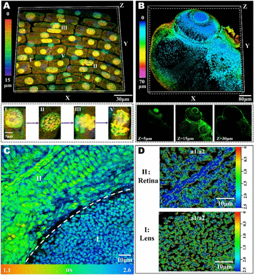
(A) 3D morphology of onion root tip apical meristem cells was stained with F-NCDs, and the images were color-coded by depth and step size: 0.5 μm. Stages I–V correspond to the interphase, prophase, metaphase, and anaphase stages of mitosis, respectively. (B) Two-photon 3D imaging of the zebrafish head, color-coded by depth, step size:1 μm. (C) FLIM imaging of zebrafish eyeballs in vivo, I: lens; II: retina. (D) a1/a2 ratio in the nucleus of lens and retina cells is color-coded by the a1/a2 ratio. |
Expression Data
Expression Detail
Antibody Labeling
Phenotype Data
Phenotype Detail
Acknowledgments
This image is the copyrighted work of the attributed author or publisher, and
ZFIN has permission only to display this image to its users.
Additional permissions should be obtained from the applicable author or publisher of the image.
Full text @ Anal. Chem.