|
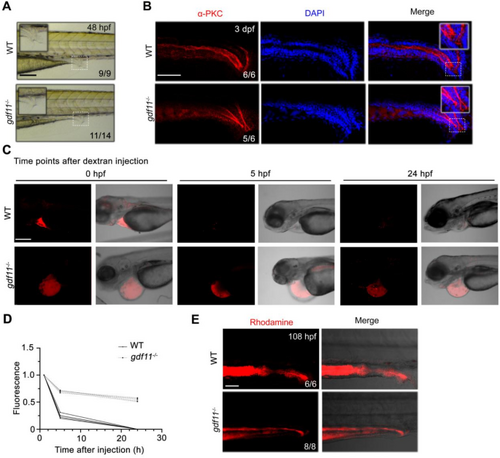
gdf11 knockout disrupts the function of the pronephros and cloaca in zebrafish. (A) Lateral view of pronephric tubules and cloaca at 36 hpf in WT and gdf11−/− mutants. Scale bar: 200 μm. (B) Lateral view of immunofluorescence images of α-PKC staining in both wild-type and gdf11 mutant embryos. Scale bar: 100 μm. (C,D) gdf11 mutant embryos exhibit slower clearance of dextran compared to WT embryos. Confocal images of embryos injected with 40 kDa rhodamine-dextran in the pericardial area immediately, and at 5–24 h post-injection (C). Quantification of fluorescence intensity (D). Scale bar: 50 μm. (E) Wild-type embryos rapidly secrete rhodamine dextran from the cloaca within minutes, while gdf11 mutants excrete only minimal dye. Scale bar: 50 μm.
|