
Fig. 1
- ID
- ZDB-FIG-250410-34
- Publication
- Olayinka et al., 2025 - Compensatory lymphangiogenesis is required for edema resolution in zebrafish
- Other Figures
- All Figure Page
- Back to All Figure Page
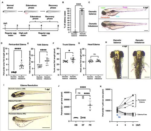
Zebrafish model of edema formation. (A) Schematic showing the experimental workflow from osmotic stress induction to edema formation and resolution. (B) Percentage of edematous larvae in osmotically balanced and osmotically imbalanced conditions at 4 dpf. Individual data points represent an independent experiment, and 20 larvae were used for each experiment. (C) Representative brightfield images showing lateral views of osmotically balanced (top) and osmotically imbalanced (bottom) 4 dpf zebrafish larvae. The dotted outline illustrates the boundary for edema area quantification in different parts of the body. Casper fish were used for imaging. (D–G) Quantification of pericardial area (D), yolk area (E), trunk area (F), and (G) head area. (H) Representative brightfield images showing the dorsal view of osmotically balanced (left) and osmotically imbalanced (right) zebrafish larvae. Red arrows indicate edema in the lateral side of the yolk area. (I) Representative images of osmotic balance (OB), edema-free (EF), and persistent-edema (PE) larva at 8 dpf. Casper fish were used for imaging. (J) Quantitative analysis of the pericardium and gut areas. Each dot represents an individual larva. (K) Line graph showing edema progression from 4–6 dpf. Each line represents the progressive change in the pericardial + yolk sac surface area of individual larvae over the experiment time course. The blue line is the larva raised in an osmotically balanced solution as a control. Scale bar = 500 μm (C,H,I). ns, not significant; *p < 0.05; ****p < 0.0001. |