|
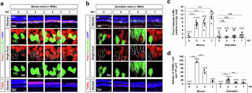
Injury-induced accumulation of Prox1 in mouse MG. a, b Retinal injury was induced in P30 mice (a) and 6-month-old zebrafish (b) through intraperitoneal injection of MNU, followed by immunostaining to assess Prox1 distribution in the retinas of both species. The bottom row displays single focal images, magnified from the boxed areas in the stacked images shown in the top row. Sox2-positive MG nuclei are outlined by dotted lines. Nuclei of the retinal cells are visualized by DAPI staining. c Relative Prox1 immunofluorescent intensity in MG at the indicated days post-injury (dpi), normalized to Prox1 intensity in BCs within the same image, is shown in the graph. Each dot represents the median intensity collected from one retina. Numbers of samples analyzed are shown in the graph (data from 4–6 independent litters). d Quantification of TUNEL-positive apoptotic cells in the specified retinal area. Columns represent mean values. Number of samples analyzed is 4. Statistical significance (p-values) of the data in (c) and (d) was calculated using one-sided Student’s t-test (*, p < 0.05; **, p < 0.01; ***, p < 0.005; ****, p < 0.001; not significant (n.s.), p > 0.05).
|